Review
. 2018 May 8:2018:5056279.
doi: 10.1155/2018/5056279. eCollection 2018.

Guiding Lights in Genome Editing for Inherited Retinal Disorders: Implications for Gene and Cell Therapy

Affiliations
Review

Guiding Lights in Genome Editing for Inherited Retinal Disorders: Implications for Gene and Cell Therapy

Carla Sanjurjo-Soriano et al. Neural Plast. .

Abstract

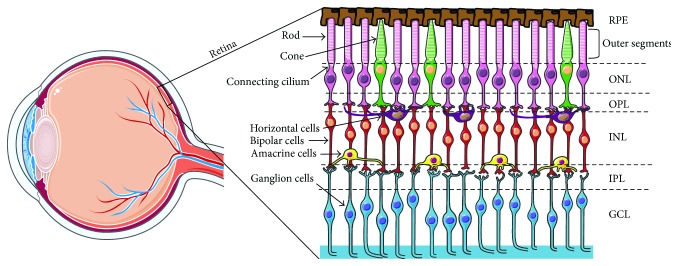
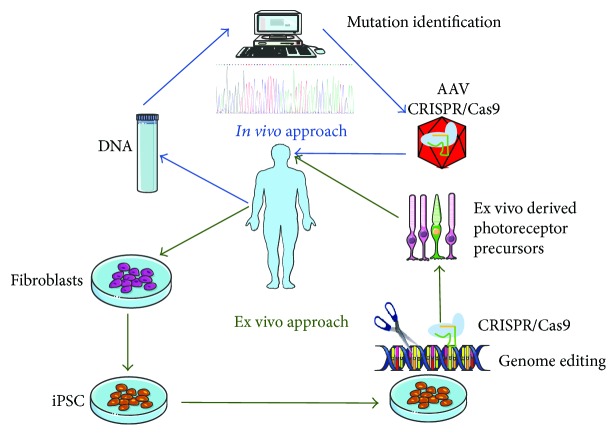
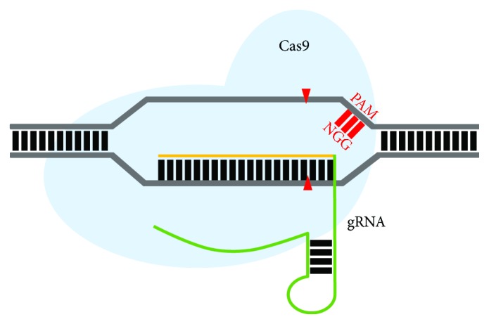
Inherited retinal dystrophies (IRDs) are a leading cause of visual impairment in the developing world. These conditions present an irreversible dysfunction or loss of neural retinal cells, which significantly impacts quality of life. Due to the anatomical accessibility and immunoprivileged status of the eye, ophthalmological research has been at the forefront of innovative and advanced gene- and cell-based therapies, both of which represent great potential as therapeutic treatments for IRD patients. However, due to a genetic and clinical heterogeneity, certain IRDs are not candidates for these approaches. New advances in the field of genome editing using Clustered Regularly Interspaced Short Palindromic Repeats (CRISPR) and CRISPR-associated protein (Cas) have provided an accurate and efficient way to edit the human genome and represent an appealing alternative for treating IRDs. We provide a brief update on current gene augmentation therapies for retinal dystrophies. Furthermore, we discuss recent advances in the field of genome editing and stem cell technologies, which together enable precise and personalized therapies for patients. Lastly, we highlight current technological limitations and barriers that need to be overcome before this technology can become a viable treatment option for patients.


Figures

Figure 1
Figure 1
Schematic representation of the retina and the retinal cell layers. The retina is a layered structure lining the back of the eye consisting of a pigmented layer, the RPE, and a multilayered neuroretina. The RPE is in close contact with the outer segments of the photosensitive rod and cone cells of the neuroretina. The connecting cilium connects the photoreceptor outer segments with the cell bodies, which constitute a layer known as the outer nuclear layer (ONL). The axons of the photoreceptors synapse with the neuronal (bipolar, amacrine, and horizontal) cells of the inner nuclear layer (INL) via the outer plexiform layer (OPL). The axons of the INL cells in turn synapse with the ganglion cell layer (GCL) via the inner plexiform layer (IPL). The axons of the ganglion cells converge to form the optic nerve.
Figure 2
Figure 2
Therapeutic approaches for treating retinal dystrophies. For an in vivo approach (indicated in blue), the patient's DNA is isolated, and genetic screening is carried out to identify the pathogenic mutation causing the retinal phenotype. Delivery of the CRISPR/Cas9 components to correct the pathogenic mutation in vivo is achieved via AAV vectors administrated directly to the retina of the patients. For an ex vivo approach (in green), patient's fibroblasts with a known mutation in an IRD gene are isolated and reprogrammed to patient-specific iPSC. Genome editing of iPSCs is carried out using the CRISPR/Cas9 system. The corrected iPSCs are further differentiated into retinal cells, which can then be reimplanted into the patient's retina.
Figure 3
Figure 3
Schematic representation of a double-stranded break (DSB; red arrowheads), which can be repaired through nonhomologous end-joining (NHEJ) or homology-directed repair (HDR) pathways. The introduction of a double-strand break in the DNA will typically undergo the error-prone NHEJ repair pathway, which results in insertions and deletions (INDELs) of variable length that will lead to premature stop codon formation. HDR, an error-free repair pathway, occurs using a wild-type donor template with homology to the target site, which serves as a template for precise gene correction of the host's DNA.
Figure 4
Figure 4
Schematic representation of the structure of a zinc finger nuclease (ZFN) and transcription activator-like effector nucleases (TALENs). (a) Cartoon of a ZFN dimer bound to DNA. ZFNs consist of two functional domains. A DNA-binding domain composed of three zinc finger modules, each one recognizing a unique triplet (3 bp) in the DNA. The DNA-cleaving domain composed of the FokI nuclease is attached to the zinc finger modules and induces the DSB in the DNA. (b) Cartoon of a TALEN dimer bound to DNA. TALENS bind DNA using the TAL effector recognizing individual nucleotides forming the DNA-binding domain. In addition, a DNA-cleaving domain comprised of the FokI nuclease is also present and will induce the DSB at the precise location in the DNA.
Figure 5
Figure 5
Schematic representation of the CRISPR/Cas9 system. The Streptococcus pyogenes Cas9 nuclease, with a “NGG” protospacer adjacent motif (PAM) sequence, has been targeted to a 20-nucleotide guide sequence in a specific region in the genome (yellow). The gRNA is complementary to the non-PAM strand. The green line represents the gRNA scaffold, which complexes with the Cas9 nuclease (light blue) and directs it to the desired site to induce a DSB (red arrowheads) in the DNA. Cas9 mediates the DSB 3 bp upstream of the PAM sequence.

References

    1. London A., Benhar I., Schwartz M. The retina as a window to the brain—from eye research to CNS disorders. 2013;9(1):44–53. doi: 10.1038/nrneurol.2012.227. - DOI - PubMed
    1. Streilein J. W. Ocular immune privilege: therapeutic opportunities from an experiment of nature. 2003;3(11):879–889. doi: 10.1038/nri1224. - DOI - PubMed
    1. Benhar I., London A., Schwartz M. The privileged immunity of immune privileged organs: the case of the eye. 2012;3:p. 296. doi: 10.3389/fimmu.2012.00296. - DOI - PMC - PubMed
    1. Kaur C., Foulds W., Ling E. Blood–retinal barrier in hypoxic ischaemic conditions: basic concepts, clinical features and management. 2008;27(6):622–647. doi: 10.1016/j.preteyeres.2008.09.003. - DOI - PubMed
    1. Benowitz L., Yin Y. Rewiring the injured CNS: lessons from the optic nerve. 2008;209(2):389–398. doi: 10.1016/j.expneurol.2007.05.025. - DOI - PMC - PubMed

MeSH terms