. 2018 May 2:2018:9354207.
doi: 10.1155/2018/9354207. eCollection 2018.

Functional Brain Connectivity during Multiple Motor Imagery Tasks in Spinal Cord Injury

Affiliations

Functional Brain Connectivity during Multiple Motor Imagery Tasks in Spinal Cord Injury

Alkinoos Athanasiou et al. Neural Plast. .

Abstract

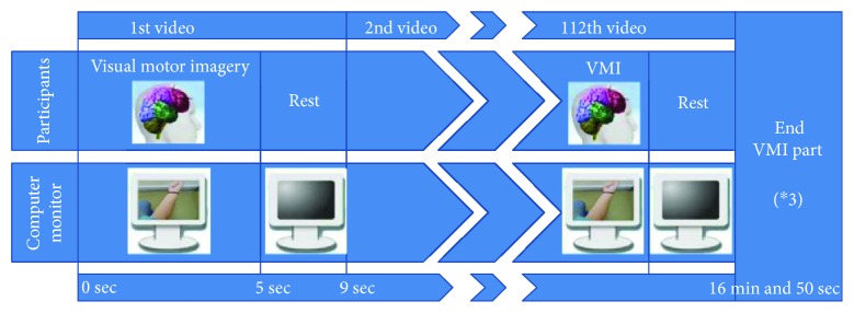
Reciprocal communication of the central and peripheral nervous systems is compromised during spinal cord injury due to neurotrauma of ascending and descending pathways. Changes in brain organization after spinal cord injury have been associated with differences in prognosis. Changes in functional connectivity may also serve as injury biomarkers. Most studies on functional connectivity have focused on chronic complete injury or resting-state condition. In our study, ten right-handed patients with incomplete spinal cord injury and ten age- and gender-matched healthy controls performed multiple visual motor imagery tasks of upper extremities and walking under high-resolution electroencephalography recording. Directed transfer function was used to study connectivity at the cortical source space between sensorimotor nodes. Chronic disruption of reciprocal communication in incomplete injury could result in permanent significant decrease of connectivity in a subset of the sensorimotor network, regardless of positive or negative neurological outcome. Cingulate motor areas consistently contributed the larger outflow (right) and received the higher inflow (left) among all nodes, across all motor imagery categories, in both groups. Injured subjects had higher outflow from left cingulate than healthy subjects and higher inflow in right cingulate than healthy subjects. Alpha networks were less dense, showing less integration and more segregation than beta networks. Spinal cord injury patients showed signs of increased local processing as adaptive mechanism. This trial is registered with NCT02443558.


Figures

Figure 1
Figure 1
Flow diagram of the experimental procedure of one part of the visual motor imagery presentation. Each presented video lasted 5 seconds and was followed by 4 seconds of black resting screen. The videos were presented 9 times each, in a random order. The presentation was divided into three parts, lasting approximately 17 minutes each, with an intermission between them.
Figure 2
Figure 2
Regions of interest (ROIs) of the sensorimotor cortex and the overlying subset of electrodes that was used for signal analysis in our study. 1: presupplementary motor area (pSMA); 2: supplementary motor area (SMA); 3: dorsal premotor area (PMd); 4: ventral premotor area (PMv); 5: cingulate motor area (CMA); 6: primary foot motor area (M1F); 7: primary hand motor area (M1H); 8: primary lip motor area (M1L); 9: primary foot somatosensory area (S1F); 10: primary hand somatosensory area (S1H); 11: secondary somatosensory area (S2); 12: somatosensory association area (SAC).
Figure 3
Figure 3
Activation time series of all regions of interest (ROIs) of the sensorimotor cortex during a random epoch and definition of time intervals around the trigger (onset of the video presenting the motor imagery task).
Figure 4
Figure 4
Average information transfer (calculated by directed transfer function) of healthy and SCI groups calculated for alpha (left) and beta (right) rhythm networks during the early imagery time interval, for the hands motor imagery category. Connections between the bilateral cingulate motor areas (CMA_R ← → CMA_L) presented the highest information transfer. Only connections with at least 25% of max information transfer among all statistically significant connections are displayed.
Figure 5
Figure 5
Nodal strengths (a: out-strength, b: in-strength) of bilateral cingulate motor areas for both subject groups. Left cingulate motor area (CMA_L) showed the highest out-strength and right cingulate motor area (CMA_R) showed the highest in-strength in the network. SCI subjects presented significantly higher CMA_R out-strength and CMA_L in-strength than healthy subjects. “∗” represent extreme values and “o” represent outliers.
Figure 6
Figure 6
Differences of group averages (healthy-SCI) of in-strength (IS) and out-strength (OS) of cingulate motor areas (CMAs) during all motor imagery categories. A trend was revealed, in which OS of CMA_R was consistently higher in the healthy than the SCI group. OS of CMA_L was consistently lower in the healthy than the SCI group. The opposite held true for IS of those nodes. This trend reached statistical significance only for early alpha walking (p = 0.002) and late beta walking (p = 0.006) tasks.
Figure 7
Figure 7
From the comparison of the networks of healthy and SCI subjects, a subset of network connections emerged as significantly stronger in the healthy group than in the SCI group for both the alpha and beta networks of “hands” motor imagery category, as calculated by network-based statistics—false discovery rate methodology.
Figure 8
Figure 8
Network density was significantly higher in beta than alpha networks (all p values < 0.001) of the “hands” motor imagery category during both early and late time intervals for all subjects of both the SCI and the healthy group (all p values < 0.001). This finding was also consistent across every other motor imagery categories in both groups (all p values < 0.001).

References

    1. Anderson K. D., Acuff M. E., Arp B. G., et al. United States (US) multi-center study to assess the validity and reliability of the spinal cord independence measure (SCIM III) 2011;49(8):880–885. doi: 10.1038/sc.2011.20. - DOI - PubMed
    1. Nardone R., Höller Y., Brigo F., et al. Descending motor pathways and cortical physiology after spinal cord injury assessed by transcranial magnetic stimulation: a systematic review. 2015;1619:139–154. doi: 10.1016/j.brainres.2014.09.036. - DOI - PubMed
    1. Athanasiou A., Klados M. A., Pandria N., et al. A systematic review of investigations into functional brain connectivity following spinal cord injury. 2017;11:p. 517. doi: 10.3389/fnhum.2017.00517. - DOI - PMC - PubMed
    1. Nardone R., Höller Y., Brigo F., et al. Functional brain reorganization after spinal cord injury: systematic review of animal and human studies. 2013;1504:58–73. doi: 10.1016/j.brainres.2012.12.034. - DOI - PubMed
    1. Freund P., Weiskopf N., Ashburner J., et al. MRI investigation of the sensorimotor cortex and the corticospinal tract after acute spinal cord injury: a prospective longitudinal study. 2013;12(9):873–881. doi: 10.1016/S1474-4422(13)70146-7. - DOI - PMC - PubMed

Publication types

Associated data