. 2018 Apr 30:2018:8309698.
doi: 10.1155/2018/8309698. eCollection 2018.

Nrf2 Deficiency Unmasks the Significance of Nitric Oxide Synthase Activity for Cardioprotection

Affiliations

Nrf2 Deficiency Unmasks the Significance of Nitric Oxide Synthase Activity for Cardioprotection

Ralf Erkens et al. Oxid Med Cell Longev. .

Abstract

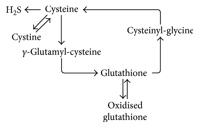
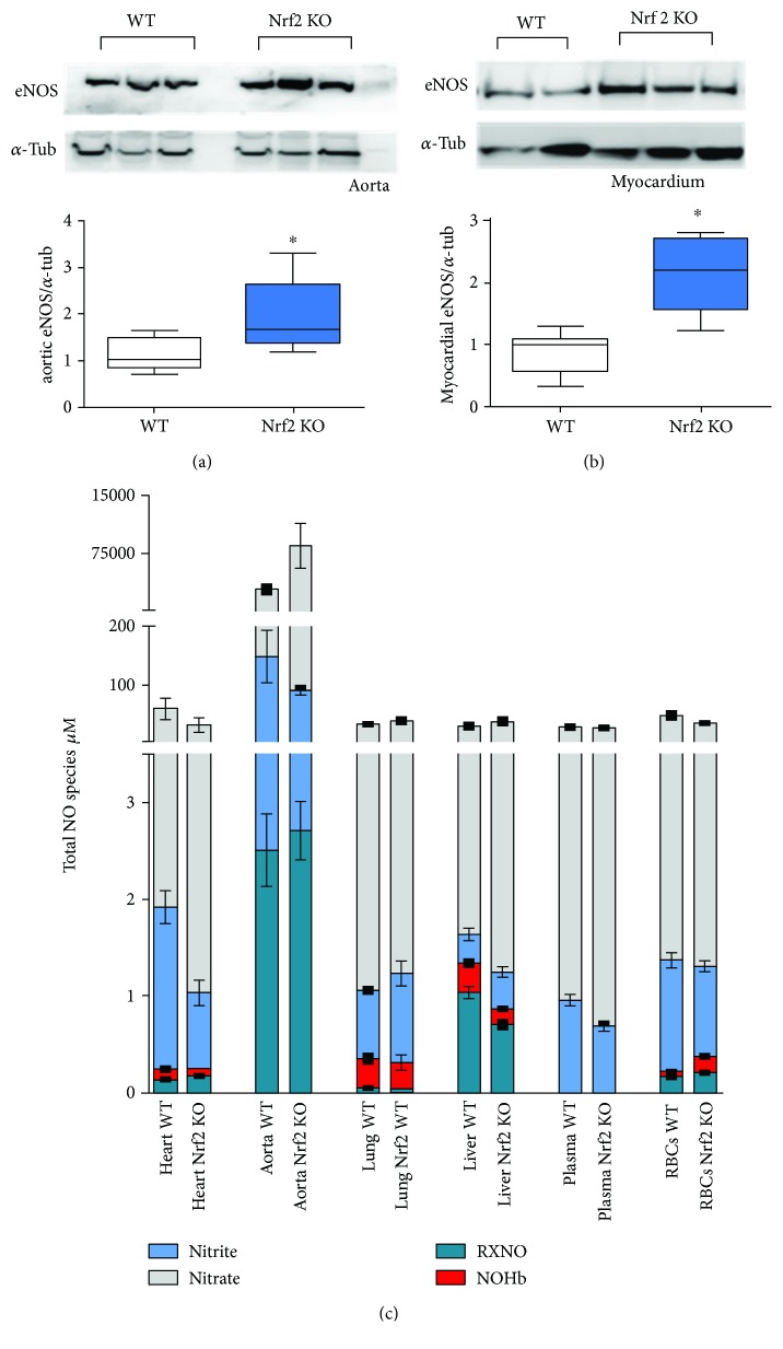
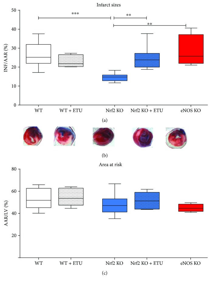
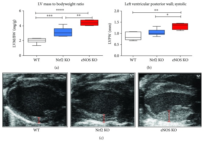
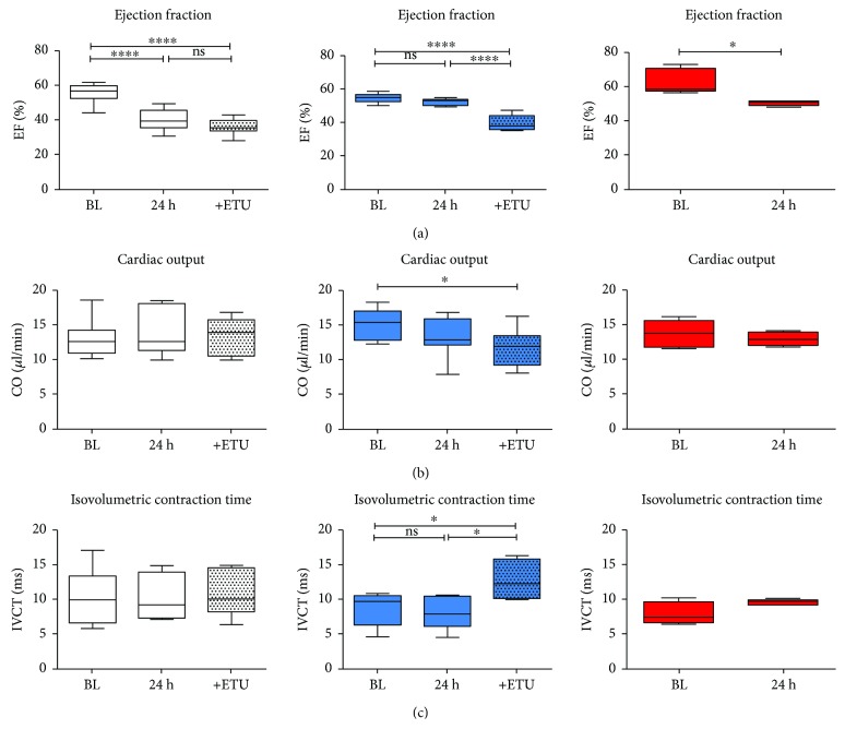
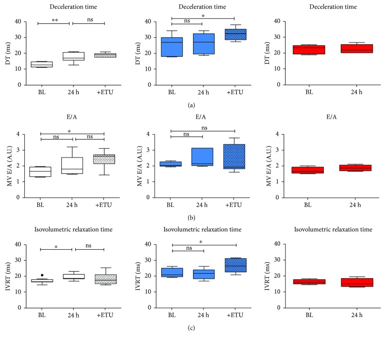
The transcription factor nuclear factor (erythroid-derived 2)-like 2 (Nrf2) is a key master switch that controls the expression of antioxidant and cytoprotective enzymes, including enzymes catalyzing glutathione de novo synthesis. In this study, we aimed to analyze whether Nrf2 deficiency influences antioxidative capacity, redox state, NO metabolites, and outcome of myocardial ischemia reperfusion (I/R) injury. In Nrf2 knockout (Nrf2 KO) mice, we found elevated eNOS expression and preserved NO metabolite concentrations in the aorta and heart as compared to wild types (WT). Unexpectedly, Nrf2 KO mice have a smaller infarct size following myocardial ischemia/reperfusion injury than WT mice and show fully preserved left ventricular systolic function. Inhibition of NO synthesis at onset of ischemia and during early reperfusion increased myocardial damage and systolic dysfunction in Nrf2 KO mice, but not in WT mice. Consistent with this, infarct size and diastolic function were unaffected in eNOS knockout (eNOS KO) mice after ischemia/reperfusion. Taken together, these data suggest that eNOS upregulation under conditions of decreased antioxidant capacity might play an important role in cardioprotection against I/R. Due to the redundancy in cytoprotective mechanisms, this fundamental antioxidant property of eNOS is not evident upon acute NOS inhibition in WT mice or in eNOS KO mice until Nrf2-related signaling is abrogated.


Figures

Scheme 1
Scheme 1
Simplified schematic representation of the main metabolic pathways involved in the production and degradation of glutathione in mammalian tissues.
Figure 1
Figure 1
Upregulation of eNOS and preserved total levels of NO metabolites in Nrf2 KO mice. (a) Representative Western blot of eNOS (upper panel) standardized to alpha-tubulin (lower panel) and densitometric analysis of eNOS expression in aorta and (b) heart of WT and Nrf2 KO mice detected by Western blot (n = 5 per group, means ± quartiles, p < 0.05, t-test). (c) Total NO species in different tissues of Nrf2 KO and WT mice (n = 5 per group, means ± SEM), (for statistical comparisons and SD please refer also to Table 2).
Figure 2
Figure 2
NOS-dependent decrease of infarct size in Nrf2 KO mice. (a) Infarct sizes (INF) per area at risk (AAR). Nrf2 KO mice showed a significant decrease in infarct sizes as compared to WT mice. Application of the NOS inhibitor ethylthiourea (ETU) in WT mice did not affect infarct size, whereas ETU application in Nrf2 KO mice increased I/R injury demonstrating the cardioprotective role of NOS activity despite a compromised antioxidative reserve capacity in Nrf2 KO mice. eNOS KO mice showed no differences in I/R injury as compared to WT and WT + ETU mice. WT mice: n = 9, Nrf2 KO mice: n = 8, eNOS KO mice: n = 4‐5; Browne-Forsythe test p = 0.27, means ± quartiles, one-way ANOVA ∗∗ p < 0.01, ∗∗∗ p < 0.001. (b) Representative TTC stained heart sections of each strain 24 h after AMI. (c) Comparable values of area at risk/left ventricle demonstrate the reproducibility of the I/R surgery (Browne-Forsythe test p = 0.24, means ± quartiles, one-way ANOVA, ns).
Figure 3
Figure 3
Cardiac hypertrophy in Nrf2 KO and eNOS KO mice. (a) LV mass normalized to body weight illustrates cardiac hypertrophy in both Nrf2 KO and eNOS KO mice as compared to WT mice. (b) Diameter of left ventricular posterior wall measured at the end of systole highlights the increase in myocardial hypertrophy in both KO mice. (c) Representative B-mode pictures of the heart from different mouse strains visualize the extent of cardiac hypertrophy. Red bars show the diameter of the left ventricular posterior wall. WT mice: n = 8, Nrf2 KO mice: n = 9, eNOS KO mice: n = 4; Browne-Forsythe test p > 0.05, means ± quartiles, one-way ANOVA p < 0.05, ∗∗ p < 0.01, ∗∗∗ p < 0.001, ∗∗∗∗ p < 0.0001.
Figure 4
Figure 4
Left ventricular systolic function following AMI in Nrf2 KO mice is preserved by NOS. (a) In WT (white box plots) and eNOS KO (red box plots) mice, ejection fraction (EF) was significantly decreased 24 h after AMI, whereas it was essentially unchanged in Nrf2 KO mice (blue box plots). ETU application resulted in an impairment of systolic function in Nrf2 KO mice, but not in WT mice, indicating NOS-dependent preservation of cardiac function in Nrf2 KO mice as compared to WT mice. (b) Cardiac output (CO) was significantly decreased after ETU application in Nrf2 KO mice indicating eNOS-dependent preservation of systolic function after I/R. (c) IVCT was increased after ETU application in Nrf2 KO mice, but not in WT mice. WT mice: n = 9, Nrf2 KO mice: n = 8; Browne-Forsythe test p > 0.05, means ± quartiles, one-way ANOVA, except for eNOS KO mice (red boxes): 4-5, unpaired t-test. p < 0.05 and ∗∗∗∗ p < 0.0001.
Figure 5
Figure 5
Nrf2 KO mice are protected against impairment of myocardial diastolic function following AMI. (a) I/R injury resulted in an impairment of diastolic function (evaluated with prolonged deceleration time (DT)), (b) E/A relation, and (c) relaxation time (IVRT) in WT mice (white box plots), whereas preexisting diastolic dysfunction was not aggravated in Nrf2 KO mice (blue box plots). In eNOS KO mice (A, red box plots), diastolic dysfunction evidenced by increased DT as compared to WT mice (Supplementary Table 1) was not further exacerbated after AMI. WT mice: n = 9, Nrf2 KO mice: n = 8; Browne-Forsythe test p > 0.05, means ± quartiles, one-way ANOVA p < 0.05, ∗∗ p < 0.01 except for eNOS KO mice n = 4‐5, unpaired t-test.

References

    1. Cortese-Krott M. M., Koning A., Kuhnle G. G. C., et al. The reactive species interactome: evolutionary emergence, biological significance, and opportunities for redox metabolomics and personalized medicine. 2017;27(10):684–712. doi: 10.1089/ars.2017.7083. - DOI - PMC - PubMed
    1. Xu B., Zhang J., Strom J., Lee S., Chen Q. M. Myocardial ischemic reperfusion induces de novo Nrf2 protein translation. 2014;1842(9):1638–1647. doi: 10.1016/j.bbadis.2014.06.002. - DOI - PMC - PubMed
    1. Heiss E. H., Schachner D., Werner E. R., Dirsch V. M. Active NF-E2-related factor (Nrf2) contributes to keep endothelial NO synthase (eNOS) in the coupled state role of reactive oxygen species (ROS), eNOS, and heme oxygenase (HO-1) levels. 2009;284(46):31579–31586. doi: 10.1074/jbc.M109.009175. - DOI - PMC - PubMed
    1. Ishii T., Itoh K., Takahashi S., et al. Transcription factor Nrf2 coordinately regulates a group of oxidative stress-inducible genes in macrophages. 2000;275(21):16023–16029. doi: 10.1074/jbc.275.21.16023. - DOI - PubMed
    1. Itoh K., Chiba T., Takahashi S., et al. An Nrf2/small Maf heterodimer mediates the induction of phase II detoxifying enzyme genes through antioxidant response elements. 1997;236(2):313–322. doi: 10.1006/bbrc.1997.6943. - DOI - PubMed

MeSH terms

LinkOut - more resources