. 2018 May 2:2018:9314626.
doi: 10.1155/2018/9314626. eCollection 2018.

Actuator-Assisted Calibration of Freehand 3D Ultrasound System

Affiliations

Actuator-Assisted Calibration of Freehand 3D Ultrasound System

Terry K Koo et al. J Healthc Eng. .

Abstract

Freehand three-dimensional (3D) ultrasound has been used independently of other technologies to analyze complex geometries or registered with other imaging modalities to aid surgical and radiotherapy planning. A fundamental requirement for all freehand 3D ultrasound systems is probe calibration. The purpose of this study was to develop an actuator-assisted approach to facilitate freehand 3D ultrasound calibration using point-based phantoms. We modified the mathematical formulation of the calibration problem to eliminate the need of imaging the point targets at different viewing angles and developed an actuator-assisted approach/setup to facilitate quick and consistent collection of point targets spanning the entire image field of view. The actuator-assisted approach was applied to a commonly used cross wire phantom as well as two custom-made point-based phantoms (original and modified), each containing 7 collinear point targets, and compared the results with the traditional freehand cross wire phantom calibration in terms of calibration reproducibility, point reconstruction precision, point reconstruction accuracy, distance reconstruction accuracy, and data acquisition time. Results demonstrated that the actuator-assisted single cross wire phantom calibration significantly improved the calibration reproducibility and offered similar point reconstruction precision, point reconstruction accuracy, distance reconstruction accuracy, and data acquisition time with respect to the freehand cross wire phantom calibration. On the other hand, the actuator-assisted modified "collinear point target" phantom calibration offered similar precision and accuracy when compared to the freehand cross wire phantom calibration, but it reduced the data acquisition time by 57%. It appears that both actuator-assisted cross wire phantom and modified collinear point target phantom calibration approaches are viable options for freehand 3D ultrasound calibration.


Figures

Figure 1
Figure 1
Principle of freehand 3D ultrasound. Each ultrasound pixel of an ultrasound image (vI) can be expressed with respect to the world coordinate system (vW) by multiplying two transformation matrixes (WTP and PTI): vW = WTP · PTI · vI, where PTI and WTP are 4 × 4 transformation matrix relating the image coordinate system to the probe coordinate system and the probe coordinate system to the world coordinate system, respectively. Although WTP can be determined from the tracking system, PTI needs to be obtained through probe calibration.
Figure 2
Figure 2
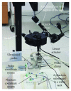
The setup for implementing the actuator-assisted calibration.
Figure 3
Figure 3
Ultrasound images of a collinear point target phantom after the one-time alignment process of the point targets. The top left image was captured with the linear actuator fully extended. The arrow indicates the lowest point of the footprint of the ultrasound probe, which is ~10 mm above the point targets (i.e., between the two parallel lines). The probe was retracted at 10 mm increment to cover the entire image field of view at 6 depth levels. Due to the curvilinear nature of the ultrasound probe used in this study, all of the 7 point targets can be clearly visualized at each depth level except for the most superficial level; only 5 screw heads can be visualized.
Figure 4
Figure 4
(a) The original collinear point target phantom; (b) the modified collinear point target phantom. The top plate of the modified collinear point target phantom is slightly rotated about the central screw to better illustrate the phantom design.
Figure 5
Figure 5
Variation of (a) translational and (b) rotational calibration parameters among the 5 calibration approaches evaluated in this study.

References

    1. Barber L., Barrett R., Lichtwark G. Validation of a freehand 3D ultrasound system for morphological measures of the medial gastrocnemius muscle. 2009;42(9):1313–1319. doi: 10.1016/j.jbiomech.2009.03.005. - DOI - PubMed
    1. Fenster A., Downey D. B., Cardinal H. N. Three-dimensional ultrasound imaging. 2001;46(5):R67–R99. doi: 10.1088/0031-9155/46/5/201. - DOI - PubMed
    1. Koo T. K., Guo J. Y., Ippolito C., Bedle J. C. Assessment of scoliotic deformity using spinous processes: comparison of different analysis methods of an ultrasonographic system. 2014;37(9):667–677. doi: 10.1016/j.jmpt.2014.09.007. - DOI - PubMed
    1. Koo T. K., Kwok W. E. A non-ionizing technique for three-dimensional measurement of the lumbar spine. 2016;49(16):4073–4079. doi: 10.1016/j.jbiomech.2016.10.048. - DOI - PubMed
    1. Kaspersen J. H., Sjolie E., Wesche J., et al. Three-dimensional ultrasound-based navigation combined with preoperative CT during abdominal interventions: a feasibility study. 2003;26(4):347–356. doi: 10.1007/s00270-003-2690-1. - DOI - PubMed

Publication types