

Fetal Midgut Volvulus with Meconium Peritonitis Detected on Prenatal Ultrasound
- PMID: 29854513
- PMCID: PMC5960549
- DOI: 10.1155/2018/5312179
Fetal Midgut Volvulus with Meconium Peritonitis Detected on Prenatal Ultrasound
Abstract
Background: Fetal volvulus is a rare, yet life-threatening condition that requires skilful diagnosis and management. Volvulus occurs when bowel loops become twisted and the twisting of the mesenteric artery leads to congestion, impaired venous return, and bowel necrosis.
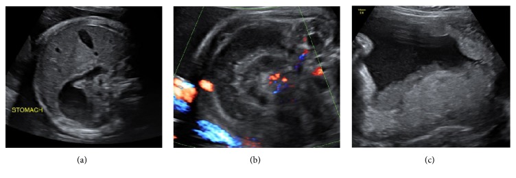
Case description: We present a case of fetal ileal volvulus suspected on third trimester ultrasound, complicated by premature labour, small bowel necrosis, and meconium peritonitis. Progressive dilatation and decreased peristalsis of echogenic bowel were noted in the early part of the third trimester. Daily surveillance ultrasound was performed and spontaneous labour occurred at 32 weeks' gestation. A proactive postnatal approach guided by prenatal sonographic findings allowed prompt treatment and an urgent laparotomy was performed for an ileal volvulus with necrosis and meconium peritonitis. A segment of small bowel volvulus was resected and an end-to-end anastomosis was performed with uneventful recovery.
Discussion: Clinically signs of fetal midgut volvulus are not pathognomonic, such as intestinal dilatation, abdominal mass, ascites, peritoneal calcifications, or polyhydramnios; thus, the diagnosis is often challenging. Complications reported in the literature include perforation and haemorrhagic ascites, which may lead to anaemia, hypovolemia, heart failure, and fetal demise.
Conclusion: This case highlights the importance of assessing the fetal bowel as a part of routine third trimester ultrasound. The case describes the complexity of diagnosis in the fetus, important considerations along with multidisciplinary team approach to management.
Figures


References
-
- Stockmann P. T. 2nd. Vol. 2. Philadelphia, Penn, USA: Lippincott Williams & Wilkins; 2005. Malrotation; p. p. 1283.
-
- Best K. E., Tennant P. W., Addor M. C. Epidemiology of small intestinal atresia in Europe: A register-based study. 2012;97:p. 353. - PubMed
Publication types
LinkOut - more resources
Full Text Sources
Other Literature Sources
