. 2018 May 6:2018:3538645.
doi: 10.1155/2018/3538645. eCollection 2018.

Perfusion Imaging in Autoimmune Encephalitis

Affiliations

Perfusion Imaging in Autoimmune Encephalitis

Deepak Vallabhaneni et al. Case Rep Radiol. .

Abstract

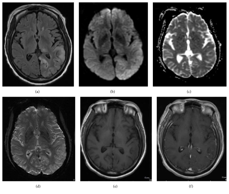
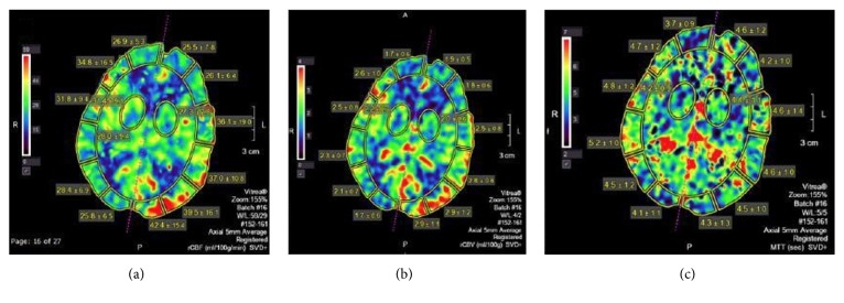
Encephalitis is characterized by inflammation of brain tissue and has various infectious and noninfectious causes. CSF analysis and MRI usually reveal inflammatory changes although sometimes brain imaging may be normal. Autoimmune encephalitis is caused by antibodies against neuronal synaptic receptors, surface proteins, or intracellular proteins. In this case report, we present a 65-year-old female who presented with a fall and altered mental status. Workup for infectious etiologies was negative and MRI of the brain displayed focal restricted diffusion with corresponding T2-FLAIR hyperintensity involving gray matter structures, making the diagnosis unclear. CT perfusion of the brain demonstrated increased cerebral blood volume and cerebral blood flow in the left parietooccipital gray matter, with corresponding normal mean transit time. Following treatment failure with acyclovir, antibiotics, and steroids, the patient was found to be positive for GAD65 antibodies and diagnosed with autoimmune encephalitis. Symptoms markedly improved with plasmapheresis. Autoimmune encephalitis rarely causes restricted diffusion and this is the first case report to describe corresponding hyperperfusion on CT perfusion study.


Figures

Figure 1
Figure 1
64-year-old female with confusion, right lower extremity weakness, and questionable encephalitis. Axial FLAIR MR image (a) shows increased signal intensity and gyriform swelling in the cortex and subcortical white matter of the left parietooccipital lobe and a focus in the left thalamus. Diffusion weighed (b) and attenuation diffusion coefficient, ADC, MR images (c) reveal mild decreased diffusivity. No susceptibility artifact or contrast enhancement is observed on axial susceptibility weighted image, SWI (d), and axial T1 weighted (e) and axial T1 weighted postgadolinium images (f), respectively.
Figure 2
Figure 2
CT perfusion was performed 10 days after the initial MRI imaging. The postprocessed CT perfusion maps in axial plane at the level of the left parietooccipital cortical abnormality reveal increased cerebral blood flow (CBF) and cerebral blood volume (CBV) in the affected cortex on the left as compared to the normal brain parenchyma on the right. The time to peak (TTP) is decreased.

References

    1. Armangue T., Petit-Pedrol M., Dalmau J. Autoimmune encephalitis in children. 2012;27(11):1460–1469. doi: 10.1177/0883073812448838. - DOI - PMC - PubMed
    1. Lancaster E., Martinez-Hernandez E., Dalmau J. Encephalitis and antibodies to synaptic and neuronal cell surface proteins. 2011;77(2):179–189. doi: 10.1212/wnl.0b013e318224afde. - DOI - PMC - PubMed
    1. Kalman B. Autoimmune encephalitides: A broadening field of treatable conditions. 2017;22(1):1–13. doi: 10.1097/NRL.0000000000000087. - DOI - PubMed
    1. Lancaster E. The diagnosis and treatment of autoimmune encephalitis. 2016;12(1):1–13. doi: 10.3988/jcn.2016.12.1.1. - DOI - PMC - PubMed
    1. Dalmau J., Lancaster E., Martinez-Hernandez E., Rosenfeld M. R., Balice-Gordon R. Clinical experience and laboratory investigations in patients with anti-NMDAR encephalitis. 2011;10(1):63–74. doi: 10.1016/S1474-4422(10)70253-2. - DOI - PMC - PubMed

LinkOut - more resources