

HDR brachytherapy in vivo source position verification using a 2D diode array: A Monte Carlo study
- PMID: 29855128
- PMCID: PMC6036394
- DOI: 10.1002/acm2.12360
HDR brachytherapy in vivo source position verification using a 2D diode array: A Monte Carlo study
Abstract
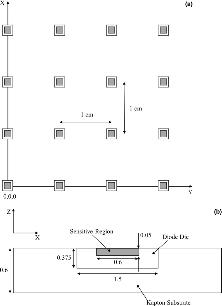
Purpose: This study aims to assess the accuracy of source position verification during high-dose rate (HDR) prostate brachytherapy using a novel, in-house developed two-dimensional (2D) diode array (the Magic Plate), embedded exactly below the patient within a carbon fiber couch. The effect of tissue inhomogeneities on source localization accuracy is examined.
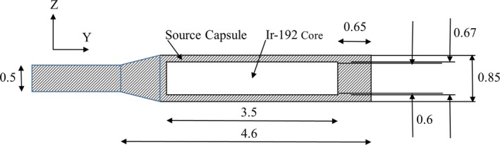
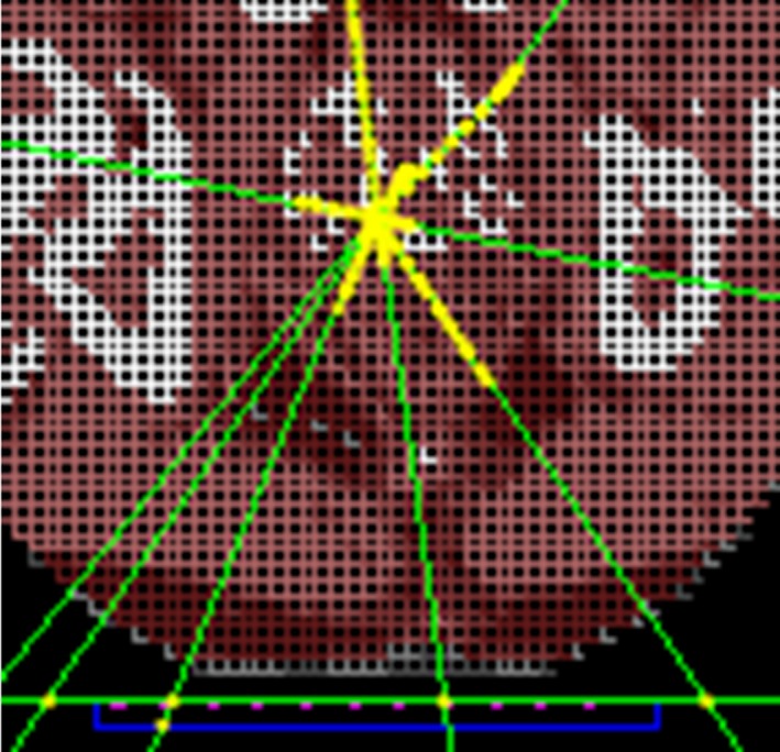
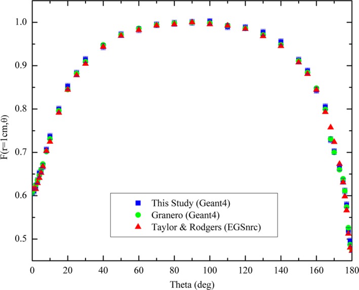
Method: Monte Carlo (MC) simulations of 12 source positions from a HDR prostate brachytherapy treatment were performed using the Geant4 toolkit. An Ir-192 Flexisource (Isodose Control, Veenendaal, the Netherlands) was simulated inside a voxelized patient geometry, and the dose deposited in each detector of the Magic Plate evaluated. The dose deposited in each detector was then used to localize the source position using a proprietary reconstruction algorithm.
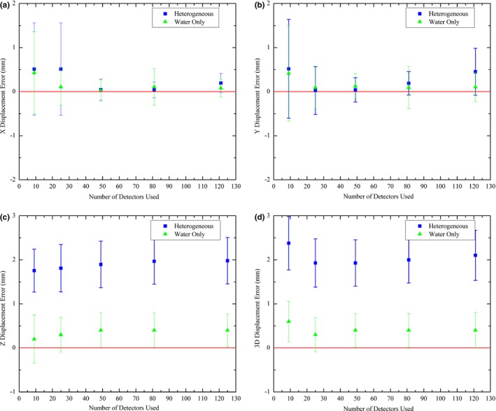
Results: The accuracy of source position verification using the Magic Plate embedded in the patient couch was found to be affected by the tissue inhomogeneities within the patient, with an average difference of 2.1 ± 0.8 mm (k = 1) between the Magic Plate predicted and known source positions. Recalculation of the simulations with all voxels assigned a density of water improved this verification accuracy to within 1 mm.
Conclusion: Source position verification using the Magic Plate during a HDR prostate brachytherapy treatment was examined using MC simulations. In a homogenous geometry (water), the Magic Plate was able to localize the source to within 1 mm, however, the verification accuracy was negatively affected by inhomogeneities; this can be corrected for by using density information obtained from CT, making the proposed tool attractive for use as a real-time in vivo quality assurance (QA) device in HDR brachytherapy for prostate cancer.
Keywords: in vivo; Magic Plate; brachytherapy; diode; source tracking.
© 2018 The Authors. Journal of Applied Clinical Medical Physics published by Wiley Periodicals, Inc. on behalf of American Association of Physicists in Medicine.
Figures
References
-
- Hoskin PJ, Rojas AM, Bownes PJ, et al. Randomised trial of external beam radiotherapy alone or combined with high‐dose‐rate brachytherapy boost for localised prostate cancer. Radiother Oncol. 2012;103:217–222. - PubMed
-
- Michalski JM, Moughan J, Purdy J, et al. A randomized trial of 79.2 Gy versus 70.2 Gy radiation therapy (RT) for localized prostate cancer. J Clin Oncol (Meeting Abstracts). 2015;33:4.
-
- Tanderup K, El Naqa I, Carlson DJ, Klein EE. Advances in Image‐Guided Brachytherapy. Int J Radiat Oncol Biol Phys. 2017;97:873–875.
-
- Tanderup K, Fokdal LU, Sturdza A, et al. Effect of tumor dose, volume and overall treatment time on local control after radiochemotherapy including MRI guided brachytherapy of locally advanced cervical cancer. Radiother Oncol. 2016;120:441–446. - PubMed
-
- Nkiwane KS, Pötter R, Tanderup K, et al. Single line source with and without vaginal loading and the impact on target coverage and organ at risk doses for cervix cancer Stages IB, II, and IIIB: treatment planning simulation in patients treated with MRI‐guided adaptive brachytherapy in a multicentre study (EMBRACE). Brachytherapy. 2013;12:317–323. - PubMed
MeSH terms
Substances
LinkOut - more resources
Full Text Sources
Other Literature Sources
