

Invasive inflammatory fibroid polyp of the stomach: a case report and literature review
- PMID: 29855265
- PMCID: PMC5984322
- DOI: 10.1186/s12876-018-0808-9
Invasive inflammatory fibroid polyp of the stomach: a case report and literature review
Abstract
Background: Inflammatory fibroid polyps (IFPs) are rare mesenchymal lesions that affect the gastrointestinal tract. IFPs are generally considered benign, noninvasive lesions; however, we report a case of an invasive gastric IFP. To the best of our knowledge, this is only the second case report of an invasive gastric IFP.
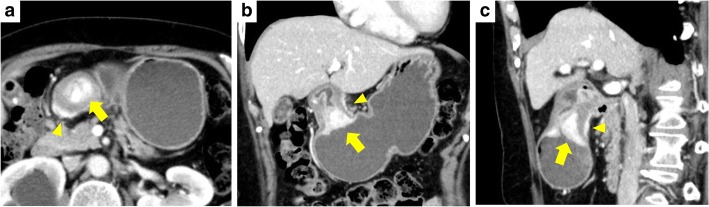
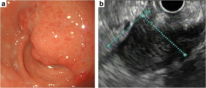
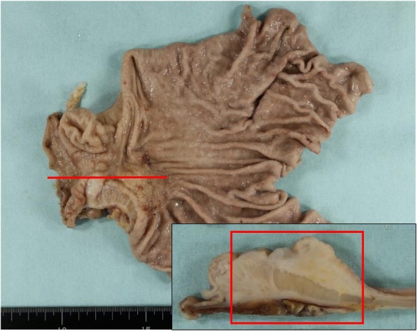
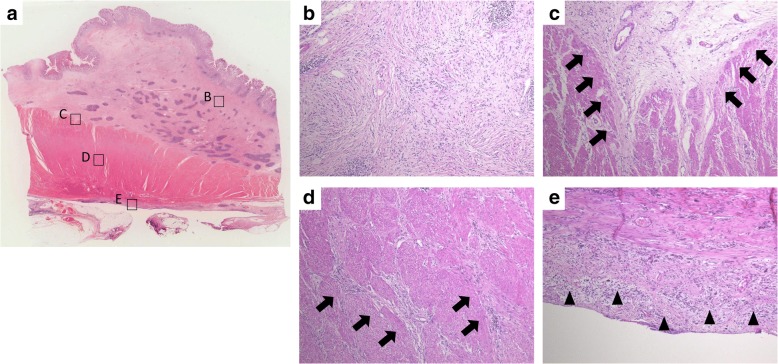
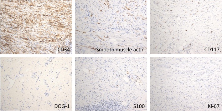
Case presentation: A 62-year-old woman presented with complaints of epigastric pain and vomiting. Computed tomography showed a 27-mm, hyper-enhancing tumor in the prepyloric antrum. Upper endoscopy also showed a submucosal tumor causing subtotal obstruction of the gastric outlet. Because a gastrointestinal stromal tumor was suspected, distal gastrectomy was performed. Histopathological examination revealed spindle cell proliferation in the submucosal layer. The spindle cells had invaded the muscularis propria layer and extended to the subserosal layer. The tumor was finally diagnosed as an IFP based on immunohistochemical findings. No mutations were identified in the platelet-derived growth factor receptor alpha (PDGFRA) gene via molecular genetic analysis.
Discussion and conclusions: After the discovery that IFPs often harbor PDGFRA mutations, these growths have been considered neoplastic lesions rather than reactive lesions. Based on the present case, IFPs might be considered not only neoplastic but also potentially invasive lesions.
Keywords: Inflammatory fibroid polyp; Invasion; Platelet-derived growth factor receptor alpha mutation; Stomach.
Conflict of interest statement
Ethics approval and consent to participate
Not applicable.
Consent for publication
Written informed consent was obtained from the patient before publication of this case report.
Competing interests
The authors declare that they have no competing interests.
Publisher’s Note
Springer Nature remains neutral with regard to jurisdictional claims in published maps and institutional affiliations.
Figures
Similar articles
-
Gastric Inflammatory Fibroid Polyp: A Rare Cause of Occult Upper Gastrointestinal Bleeding.J Investig Med High Impact Case Rep. 2020 Jan-Dec;8:2324709620936840. doi: 10.1177/2324709620936840. J Investig Med High Impact Case Rep. 2020. PMID: 32602395 Free PMC article. Review.
-
[A giant inflammatory fibroid polyp of the esophagus].Magy Seb. 2017 Mar;70(1):69-73. doi: 10.1556/1046.70.2017.1.10. Magy Seb. 2017. PMID: 28294664 Hungarian.
-
Gastric outlet obstruction due to inflammatory fibroid polyp.Ann Ital Chir. 2006 Jan-Feb;77(1):59-61. Ann Ital Chir. 2006. PMID: 16910362
-
Inflammatory fibroid polyp of the renal pelvis: first report at an extra-gastrointestinal site with molecular confirmation.Virchows Arch. 2023 Oct;483(4):535-539. doi: 10.1007/s00428-023-03557-y. Epub 2023 May 15. Virchows Arch. 2023. PMID: 37184764 Free PMC article.
-
[Inflammatory fibroid polyp: from Vanek's "submucosal granuloma" to the concept of submucosal mesenchymal neoplasia].Pathologe. 2010 Mar;31(2):109-14. doi: 10.1007/s00292-009-1254-9. Pathologe. 2010. PMID: 20107807 Review. German.
Cited by
-
Jejunal Intussusception Secondary to a Large Inflammatory Fibroid Polyp: A Case Report and Discussion of Differential Diagnosis.Case Rep Pathol. 2023 Apr 13;2023:9417141. doi: 10.1155/2023/9417141. eCollection 2023. Case Rep Pathol. 2023. PMID: 37091748 Free PMC article.
-
Comparison of endoscopic ultrasonography features and pathological staging of gastric inflammatory fibroid polyps.World J Gastrointest Surg. 2025 Jul 27;17(7):105136. doi: 10.4240/wjgs.v17.i7.105136. World J Gastrointest Surg. 2025. PMID: 40740919 Free PMC article.
-
Gastric Inflammatory Fibroid Polyp: A Rare Cause of Occult Upper Gastrointestinal Bleeding.J Investig Med High Impact Case Rep. 2020 Jan-Dec;8:2324709620936840. doi: 10.1177/2324709620936840. J Investig Med High Impact Case Rep. 2020. PMID: 32602395 Free PMC article. Review.
-
Inflammatory Fibroid Polyp of the Gastrointestinal Tract: A Systematic Review for a Benign Tumor.In Vivo. 2021 Jan-Feb;35(1):81-93. doi: 10.21873/invivo.12235. In Vivo. 2021. PMID: 33402453 Free PMC article.
-
Inflammatory fibroid polyp in the antrum co-occurring with adenomatous polyp in the ascending colon.Prz Gastroenterol. 2018;13(4):340-342. doi: 10.5114/pg.2018.79816. Epub 2018 Dec 11. Prz Gastroenterol. 2018. PMID: 30581510 Free PMC article. No abstract available.
References
Publication types
MeSH terms
LinkOut - more resources
Full Text Sources
Other Literature Sources
Medical
Miscellaneous
