. 2018 May 31;6(1):43.
doi: 10.1186/s40478-018-0543-z.

A common antigenic motif recognized by naturally occurring human VH5-51/VL4-1 anti-tau antibodies with distinct functionalities

Affiliations

A common antigenic motif recognized by naturally occurring human VH5-51/VL4-1 anti-tau antibodies with distinct functionalities

Adrian Apetri et al. Acta Neuropathol Commun. .

Abstract

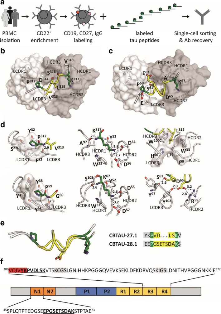
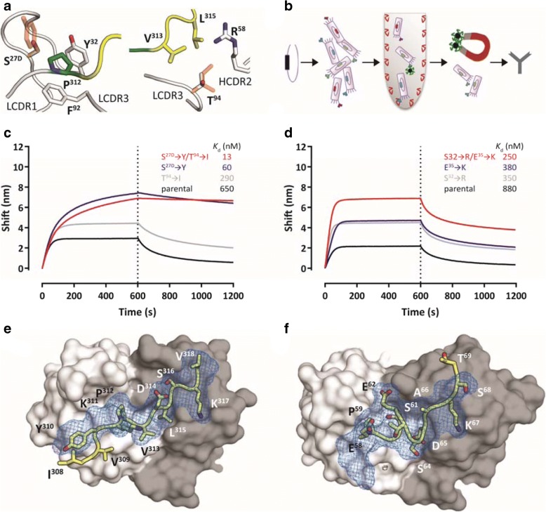
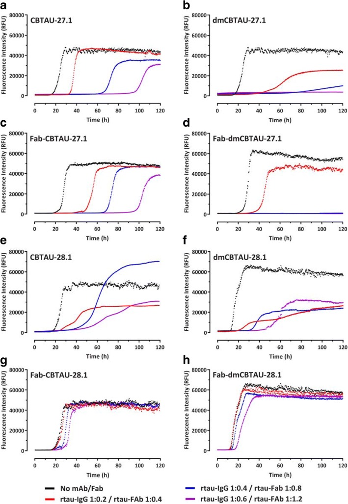
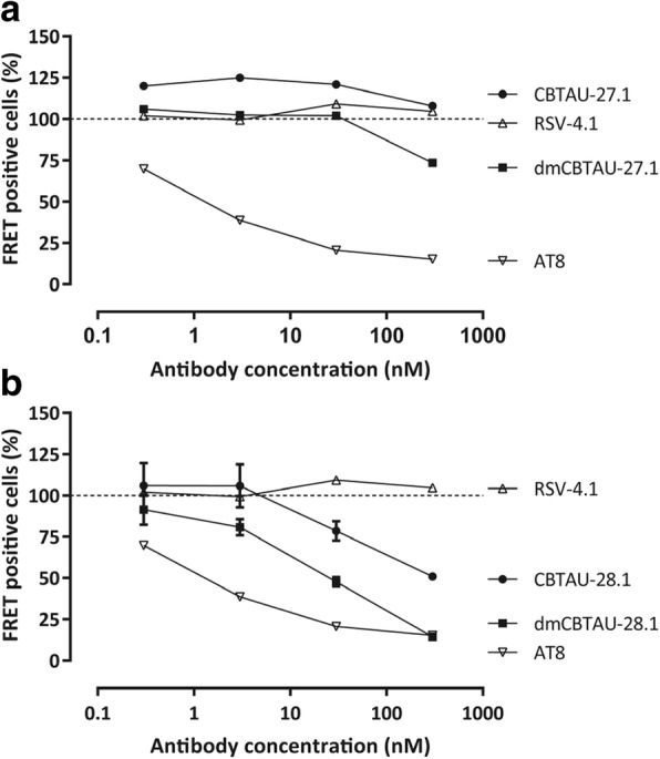
Misfolding and aggregation of tau protein are closely associated with the onset and progression of Alzheimer's Disease (AD). By interrogating IgG+ memory B cells from asymptomatic donors with tau peptides, we have identified two somatically mutated VH5-51/VL4-1 antibodies. One of these, CBTAU-27.1, binds to the aggregation motif in the R3 repeat domain and blocks the aggregation of tau into paired helical filaments (PHFs) by sequestering monomeric tau. The other, CBTAU-28.1, binds to the N-terminal insert region and inhibits the spreading of tau seeds and mediates the uptake of tau aggregates into microglia by binding PHFs. Crystal structures revealed that the combination of VH5-51 and VL4-1 recognizes a common Pro-Xn-Lys motif driven by germline-encoded hotspot interactions while the specificity and thereby functionality of the antibodies are defined by the CDR3 regions. Affinity improvement led to improvement in functionality, identifying their epitopes as new targets for therapy and prevention of AD.

Keywords: Alzheimer’s disease; Antigenic motif; Monoclonal antibody; Tau protein.


Conflict of interest statement

Ethics approval and consent to participate

Brain samples were obtained from The Netherlands Brain Bank (NBB), Netherlands Institute for Neuroscience, Amsterdam. All donors had given written informed consent for brain autopsy and the use of material and clinical information for research purposes. Whole blood from healthy male and female donors was obtained from the San Diego Blood Bank (ages 18–65 years) after informed consent was obtained from the donors.

Competing interests

The authors declare that they have no competing interests.

Publisher’s Note

Springer Nature remains neutral with regard to jurisdictional claims in published maps and institutional affiliations.

Figures

Fig. 1
Fig. 1
Recovery and structural characterization of naturally occurring monoclonal antibodies to unphosphorylated tau epitopes from asymptomatic individuals. a BSelex method used to recover tau-specific memory B cells. PBMCs were prepared from asymptomatic blood bank donors, and mature CD22+ B cells were positively selected with magnetic beads. Viable cells were stained with IgG-FITC, CD19-PerCPCy5.5, and CD27-PECy7, and with a pool of 10 overlapping unphosphorylated tau peptides spanning the longest tau isoform (relative position of each peptide along 2N4R tau indicated). All peptides were present in the pool with an APC label as well as with a PE label and CD19+, CD27+, IgG+, APC+, PE+ cells were single-cell sorted on a Beckman Coulter MoFlo XDP. Antibody heavy and light variable chain sequences were recovered from single cells, cloned and expressed as full-length IgGs. b and c Co-crystal structures of Fab CBTAU-27.1 (b) and Fab CBTAU-28.1 (c) with tau peptides A8119 and A7731, respectively. Antibodies have been plotted as molecular surface with light chain in white and heavy chain in grey. Tau peptides are shown as cartoon with interacting amino acids plotted as sticks. Proline and lysine residues are plotted in green, amino acids in between these residues are colored in yellow and the termini in grey. Only the interacting antibody loops are outlined. d Key interactions with tau of CBTAU-27.1 (upper row) and CBTAU-28.1 (lower row). Key interacting residues are plotted as sticks, polar interactions are indicated with dotted lines, and the corresponding distances are indicated in Å. In the first panel, interactions with Pro312 and Pro59 are compared where the proline binding pockets are visualized on a molecular surface. In the second panel, interactions with Lys317 and Lys67 are compared. In panel 3, interactions around Leu315 and Asp65 in the central region of the epitopes are shown. e Structural basis for recognition of the Pro – Xn – Lys epitope motif. Epitopes of CBTAU-27.1 and CBTAU-28.1 are superimposed by aligning on the proline and lysine residues. The peptides present both residues in the same spatial orientation. In the central region (yellow), hydrophobic Leu315 in CBTAU-27.1 is replaced by hydrophilic and charged Asp65 in CBTAU-28.1. f Schematic representation of tau isoform 2N4R showing the epitopes of CBTAU-27.1 and CBTAU  -28.1 (bold and underlined) and the surrounding sequences. Highlighted in grey and red are the microtubule binding motifs and the hexapeptide 306VQIVYK311 which forms the N-terminal end of the core of PHFs, respectively. N1 and N2 indicate acidic inserts, P1 and P2 indicate proline-rich domains, and R1-R4 indicate microtubule-binding repeat domains
Fig. 2
Fig. 2
Generation of affinity-improved mutants of CBTAU-27.1 and CBTAU-28.1 a Structure-based design of mutants around Pro312 (left panel) and Val313 (right panel). The tau epitope is illustrated as in Fig. 1b. Antibody loops and the key residues interacting with Pro312 and Thr94 are plotted in white. Proposed mutations are shown as orange sticks on top of the corresponding wild-type side chains. Ser27D is mutated to tyrosine (left panel) to enlarge the hydrophobic pocket of Pro312, and Thr94 is mutated to isoleucine (right panel) to fill the empty cavity surrounding Val313 and Leu315. By introducing both mutations, additional hydrophobic contacts between tau and the antibody loops could be formed, potentially resulting in a lower desolvation penalty and increased affinity. b Schematic representation of the CBTAU-28.1 affinity maturation process by random mutagenesis. Mutations were introduced randomly by error prone PCR in the coding sequence for the single-chain variable fragment (scFv) directed against the CBTAU-28.1 epitope. M13 phage libraries displaying the scFv were screened against rtau and peptide A6940. Affinity-matured variants were identified by phage ELISA and converted into an IgG1 format to assess binding in solution. c and d Association and dissociation profiles for parental and affinity improved CBTAU-27.1 (c) and CBTAU-28.1 (d) variants to their corresponding cognate peptides as determined by Octet biolayer interferometry. Affinities as determined by ITC (Kd) are shown on the individual graphs. (e and f) Co-crystal structures of the Fabs of dmCBTAU-27.1 (e) and dmCBTAU-28.1 (f) with tau peptides A8119 and A7731, respectively. Antibodies are illustrated as molecular surfaces (colored as in panel A), together with tau epitopes as sticks with yellow carbons. The corresponding parental co-crystal structures have been aligned using their variable regions, and their tau epitopes are shown as blue mesh on top of the mutant epitopes.
Fig. 3
Fig. 3
Detection of immunoreactivity in human brain tissue by CBTAU-27.1 and CBTAU-28.1 and affinity-improved variants. Immunohistochemistry was performed on 5 μm thick formalin-fixed paraffin embedded sections of the hippocampal region using a 0.1 μg/ml antibody concentration. Immunodetection using CBTAU-27.1 (a-d), dmCBTAU-27.1 (e-h), CBTAU-28.1 (i-l), dmCBTAU-28.1 (m-p) and PHF-tau-specific mouse antibody AT8 (q-t) in control and AD brain tissue without or with heat pretreatment using sodium citrate buffer. Gallyas staining for detection of NFTs and neuropil threads is shown from the same control and AD case of corresponding areas for comparison (u, v). Immunoreactivity was visualized using DAB (brown) and nuclei were counterstained with haematoxylin (blue). Representative areas of the CA1/subiculum of the hippocampus are shown. Scale bars represent 50 μm
Fig. 4
Fig. 4
CBTAU-27.1, but not CBTAU-28.1, inhibits the aggregation of recombinant tau in vitro. Aggregation of rtau in the absence (black) or presence of CBTAU-27.1 (a), dmCBTAU-27.1 (b), Fab CBTAU-27.1 (c), Fab dmCBTAU-27.1 (d), CBTAU-28.1 (e), dmCBTAU-28.1 (f), Fab CBTAU-28.1 (g) or Fab dmCBTAU-28.1 (h), as monitored continuously for 120 h by ThT fluorescence. Three different rtau-IgG (1:0.2 – red, 1:0.4 – blue, and 1: 0.6 – purple) and rtau-Fab (1:0.4 – red, 1: 0.8 – blue, and 1:1.2 – purple) stoichiometries were tested. Each condition was tested in quadruplicate and one representative curve is shown for each condition. For complete datasets, see Additional file 1: Figures S7-S10
Fig. 5
Fig. 5
CBTAU-28.1, but not CBTAU-27.1, is capable of immunodepleting seeds from AD brains. Residual seeding activity of human AD brain homogenates following immunodepletion with different concentrations of CBTAU-27.1 and dmCBTAU-27.1 (a) or CBTAU-28.1 and dmCBTAU-28.1 (b) as measured by FRET signal in biosensor cells expressing the microtubule repeat domains of tau (aa 243–375) fused either to yellow or cyan fluorescent protein. Uptake of exogenous tau aggregates into the cells results in aggregation of the tau fusion proteins, which is detected by FRET. As positive and negative controls, a human IgG1 chimeric version of murine anti-PHF antibody AT8 and anti-RSV-G antibody RSV-4.1 were taken along, respectively. For the controls, the same data are shown in plots A and B for visualization purposes. Error bars indicate the SD of two independent experiments
Fig. 6
Fig. 6
CBTAU-28.1, but not CBTAU-27.1, enhances uptake of tau aggregates into microglial BV2 cells. a and c Aggregated recombinant tau was covalently labelled with pHrodo Green dye and incubated with chimeric versions (containing mouse instead of human Fc region) of CBTAU-28.1, dmCBTAU-28.1, CBTAU27.1, dmCBTAU-27.1, their Fab fragments, a mouse IgG1 isotype control antibody (IC), or no antibody (rtau). Immunocomplexes were subsequently incubated with BV2 cells and their uptake was assessed by flow cytometry as expressed by the geometric mean fluorescent intensity. Error bars indicate the SD of two independent experiments. b and d Preformed pHrodo-Green labeled immunocomplexes of rtau with chimeric dmCBTAU-28.1 or dmCBTAU-27.1 (at a concentration of 150 nM) were incubated with BV-2 cells. After incubation, nuclei were stained with Hoechst (blue) and the acidic cellular compartment with LysoTracker Red dye and uptake of immunocomplexes was assessed by live-cell imaging. Images represent maximum intensity projections of a 20 planes Z-stack (0.5 μm planes) acquired with a 63× water immersion objective

References

    1. Afonine PV, Grosse-Kunstleve RW, Echols N, Headd JJ, Moriarty NW, Mustyakimov M, Terwilliger TC, Urzhumtsev A, Zwart PH, Adams PD. Towards automated crystallographic structure refinement with phenix.Refine. Acta Crystallogr Sect D Biol Crystallogr. 2012;68:352–367. doi: 10.1107/S0907444912001308. - DOI - PMC - PubMed
    1. Apetri AC, Vanik DL, Surewicz WK. Polymorphism at residue 129 modulates the conformational conversion of the D178N variant of human prion protein 90-231. Biochemistry. 2005;44:15880–15888. doi: 10.1021/bi051455+. - DOI - PubMed
    1. Arevalo JH, Stura EA, Taussig MJ, Wilson IA. Three-dimensional structure of an anti-steroid Fab' and progesterone-Fab' complex. J Mol Biol. 1993;231:103–118. doi: 10.1006/jmbi.1993.1260. - DOI - PubMed
    1. Billingsley ML, Kincaid RL. Regulated phosphorylation and dephosphorylation of tau protein: effects on microtubule interaction, intracellular trafficking and neurodegeneration. Biochem J. 1997;323(Pt 3):577–591. doi: 10.1042/bj3230577. - DOI - PMC - PubMed
    1. Braak H, Braak E. Neuropathological stageing of Alzheimer-related changes. Acta Neuropathol. 1991;82:239–259. doi: 10.1007/BF00308809. - DOI - PubMed

Publication types

MeSH terms