. 2018 May 31;8(1):8463.
doi: 10.1038/s41598-018-26745-2.

Direct phenotypic conversion of human fibroblasts into functional osteoblasts triggered by a blockade of the transforming growth factor-β signal

Affiliations

Direct phenotypic conversion of human fibroblasts into functional osteoblasts triggered by a blockade of the transforming growth factor-β signal

Kenta Yamamoto et al. Sci Rep. .

Abstract

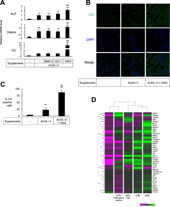
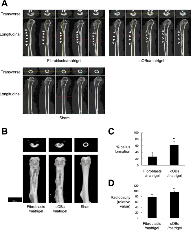
A procedure to generate functional osteoblasts from human somatic cells may pave the way to a novel and effective transplantation therapy in bone disorders. Here, we report that human fibroblasts were induced to show osteoblast phenotypes by culturing with ALK5 i II, which is a specific inhibitor for activin-like kinase 5 (ALK5) (tumor growth factor-β receptor 1 (TGF-β R1)). Cells cultured with ALK5 i II expressed osteoblast-specific genes and massively produced calcified bone matrix, similar to the osteoblasts induced from mesenchymal stem cells (MSC-OBs). Treatment with vitamin D3 in addition to ALK5 i II induced more osteoblast-like characters, and the efficiency of the conversion reached approximately 90%. The chemical compound-mediated directly converted osteoblasts (cOBs) were similar to human primary osteoblasts in terms of expression profiles of osteoblast-related genes. The cOBs abundantly produced bone matrix in vivo and facilitated bone healing after they were transplanted into immunodeficient mice at an artificially induced defect lesion in femoral bone. The present procedure realizes a highly efficient direct conversion of human fibroblasts into transgene-free and highly functional osteoblasts, which might be applied in a novel strategy of bone regeneration therapy in bone diseases.


Conflict of interest statement

The authors declare no competing interests.

Figures

Figure 1
Figure 1
Osteoblast-like phenotypes were induced in human fibroblasts cultured in osteogenic medium supplemented with ALK5 i II. (A) Human dermal fibroblasts (aHDFs) were seeded into 24-well plates and cultured in the complete medium or osteogenic medium supplemented with TGF-β receptor inhibitors or rTGF-β as indicated. After culturing for 21 days, cells were stained with Alizarin Red S. Macroscopic images (Upper) and staining intensities (OD550) (Lower) are shown. (B) aHDFs and MSCs were seeded into 24-well plates (day 0) and cultured as in (A). Thirteen days later, cells were subjected to ALP staining (Bottom). On day 18, the cells were stained with Alizarin Red S. Macroscopic images (Middle) and staining intensities (OD550) are shown (Top). (C) Cells were cultured as in (B) and RNA was subjected to the real time-RT-PCR analysis on day 18. Relative mRNA levels are plotted. Magnification of the images are x 1. Values are means ± S.D. n = 3 (A and B) or 4 (C). *P < 0.05 and **P < 0.01, vs. the aHDF cultured in osteogenic medium alone. N.D., no significant difference between the indicated groups.
Figure 2
Figure 2
An addition of ALK5 i II inhibited smad2/3 signaling in fibroblasts. (A) HDFs were seeded onto 60-mm dishes, and on the next day culture supernatant was replaced by fresh complete medium with/without the indicated concentrations of TGF-β inhibitors. After culturing for 3 days, culture supernatant was replaced by fresh one, and cells were lysed 30 min later. ELISA was performed to evaluate relative ratio of phospho-smad2/3 per total smad2/3 (the value for untreated HDFs was set to 1). Values are means ± S.D. (n = 3). *P < 0.05, **P < 0.01 vs. untreated control. #P < 0.05, ##P < 0.05 vs. HDFs treated with ALK5i II at the same concentration. (B and C) HDFs were seeded onto 60-mm dishes, and on the next day culture supernatant was replaced by fresh complete or osteogenic medium with/without ALK5 i II as indicated. After culturing for 3 days, culture supernatant was replaced by fresh one, and cells were lysed 30 min later. Western blotting analyses were performed using the indicated antibodies.
Figure 3
Figure 3
HDFs were efficiently converted into OC-producing osteoblasts by culturing with ALK5 i II and VitD3. (A) HDFs were cultured in osteogenic medium supplemented with ALK5 i II and the indicated supplements. After culturing for 18 days, real time RT-PCR was performed to evaluate mRNA for the indicated gene. Values are means ± S.D. n = 4. *P < 0.05 and **P < 0.01, v.s. HDFs cultured in osteogenic medium alone. #P < 0.05 and ##P < 0.01, v.s. HDFs cultured in osteogenic medium supplemented with ALK5 i II. (B and C), HDFs were cultured in osteogenic medium supplemented with ALK5 i II and VitD3 for 18 days (cOBs). The cOBs, the HDFs cultured in osteogenic medium supplemented with ALK5 i II and the HDFs cultured in complete medium as control were stained with anti-OC antibody and DAPI. Fluorescence microscopic images (magnification was x200) (B) and proportion of OC producing cells (C) are shown. Values are means ± S.D. (n = 4). **P < 0.01. v.s. control. ##P < 0.01, v.s. HDFs cultured in osteogenic medium supplemented with ALK5 i II. (D) RNA was extracted from HDFs, HDFs cultured in the osteogenic medium for 18 days, cOBs induced as in (B), osteoblasts differentiated from MSCs (MSC-OBs), and pOBs. DNA microarray analysis was performed to evaluate mRNA for the genes encoding osteoblasts-related transcription factors, signaling proteins and soluble factors. Heat map and hierarchical clustering data are shown. The genes with increased expression are colored green, whereas those with decreased expression are colored pink as indicated in the color range. The expression level of each gene was normalized to median signal intensity.
Figure 4
Figure 4
cOBs facilitated bone repair in vivo. HDFs were cultured in osteogenic medium supplemented with ALK5 i II and VitD3 for 13 days (cOBs). The cOBs and the HDFs cultured in complete medium as control were inoculated into artificial segmental bone defect lesion in femoral diaphysis in NOG mice. Bone defect was not created in the sham-operated mice. Twenty-one days later, mice were sacrificed and μCT imaging of the femur was performed. (A and B), Longitudinal and transverse serial 100 μm slice images (A) and 3D-constructed μCT images (B) of the femurs of a representative mouse are shown. White triangles indicate bone defect lesions, while arrow heads indicate regenerated bone tissue. (C and D), The means ± S.D. of the percentages of callus formation (C) and the relative radiopacity at the bone defect region (value for the sham operated group are set to 100%) (D) are plotted (n = 4). **P < 0.01.
Figure 5
Figure 5
cOBs contributed to bone tissue regeneration in vivo. Transplantation experiment was performed as in Fig. 3. Twenty-one days after the cell inoculation, mice were sacrificed and the femur was excised. (A) Serial sections of the femur tissue were stained with Alizarin Red S (Upper) and H & E. (Lower). Scale bar = 1 mm. (B) Sections were stained with anti-human OC antibody followed by FITC-labeld secondary antibody, anti-human vimentin antibody followed by PE-labed secondary antibody, and DAPI. Optical light and fluorescence microscopic images are shown. Scale bar = 100 μm.

References

    1. Neve A, Corrado A, Cantatore FP. Osteoblast physiology in normal and pathological conditions. Cell Tissue Res. 2011;343:289–302. doi: 10.1007/s00441-010-1086-1. - DOI - PubMed
    1. Ieda M, et al. Direct reprogramming of fibroblasts into functional cardiomyocytes by defined factors. Cell. 2010;142:375–386. doi: 10.1016/j.cell.2010.07.002. - DOI - PMC - PubMed
    1. Hiramatsu K, et al. Generation of hyaline cartilaginous tissue from mouse adult dermal fibroblast culture by defined factors. J Clin Invest. 2011;121:640–657. doi: 10.1172/JCI44605. - DOI - PMC - PubMed
    1. Kim J, et al. Direct reprogramming of mouse fibroblasts to neural progenitors. Proc Natl Acad Sci USA. 2011;108:7838–7843. doi: 10.1073/pnas.1103113108. - DOI - PMC - PubMed
    1. Nizzardo M, et al. Direct reprogramming of adult somatic cells into other lineages: past evidence and future perspectives. Cell Transplant. 2013;22:921–944. doi: 10.3727/096368912X657477. - DOI - PubMed

Publication types

MeSH terms

Substances