. 2018 May 31;8(1):8512.
doi: 10.1038/s41598-018-26869-5.

3D Printed porous polyamide macrocapsule combined with alginate microcapsules for safer cell-based therapies

Affiliations

3D Printed porous polyamide macrocapsule combined with alginate microcapsules for safer cell-based therapies

Laura Saenz Del Burgo et al. Sci Rep. .

Abstract

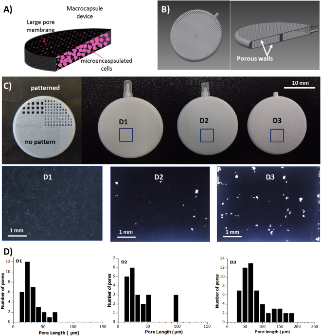
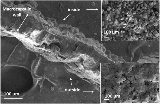
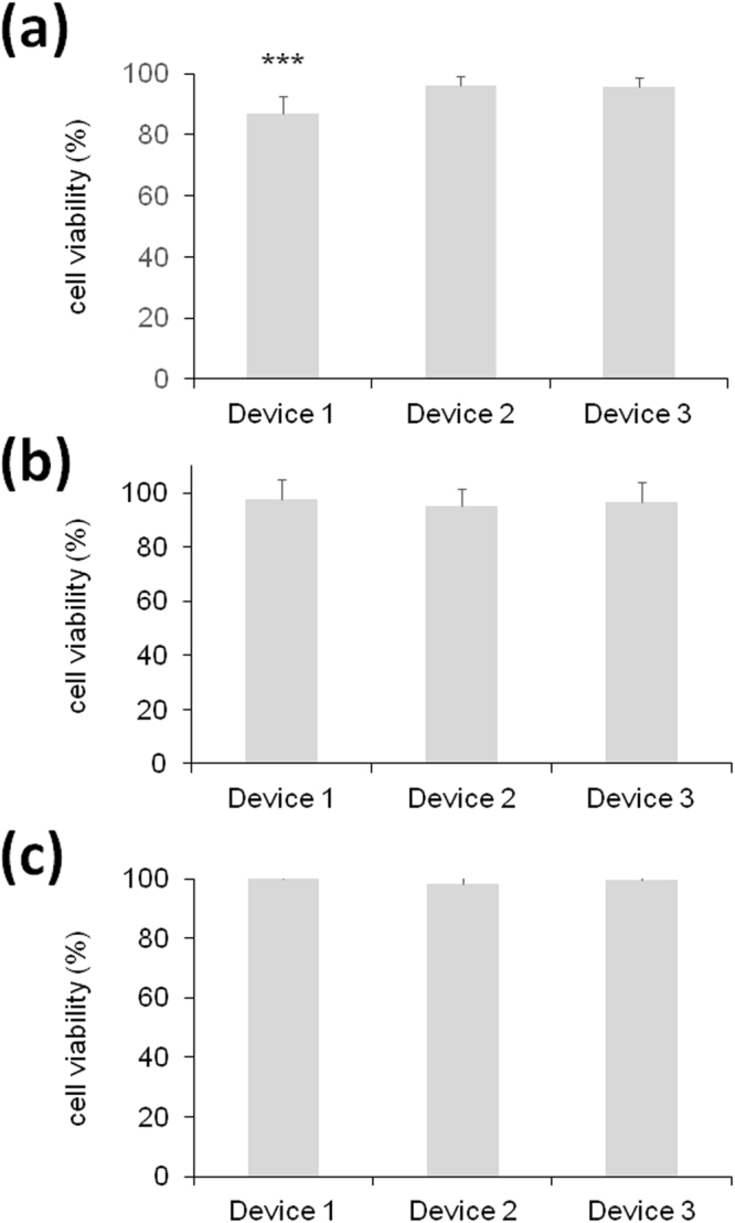
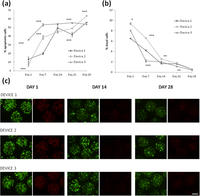
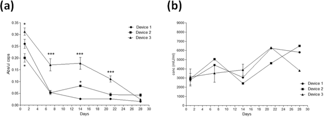
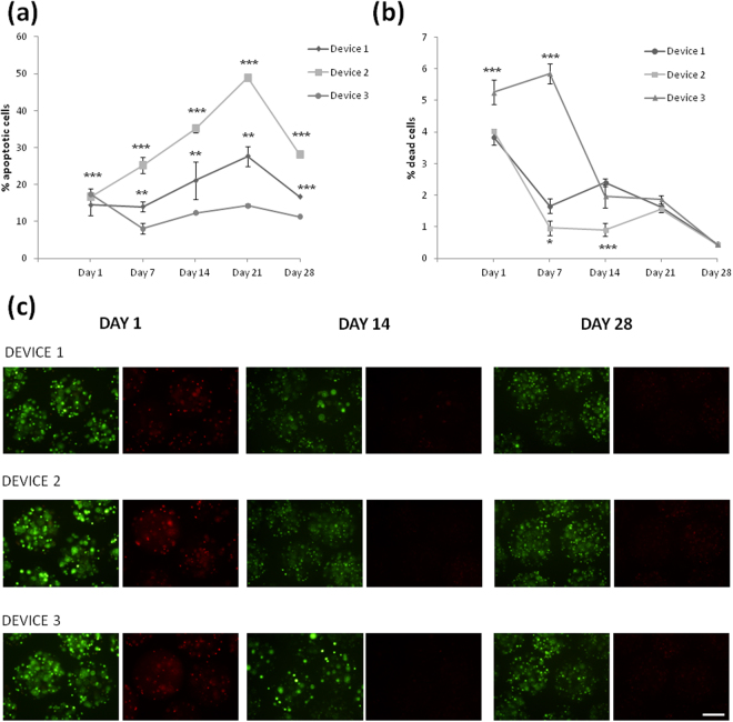
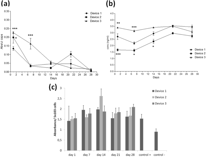
Cell microencapsulation is an attractive strategy for cell-based therapies that allows the implantation of genetically engineered cells and the continuous delivery of de novo produced therapeutic products. However, the establishment of a way to retrieve the implanted encapsulated cells in case the treatment needs to be halted or when cells need to be renewed is still a big challenge. The combination of micro and macroencapsulation approaches could provide the requirements to achieve a proper immunoisolation, while maintaining the cells localized into the body. We present the development and characterization of a porous implantable macrocapsule device for the loading of microencapsulated cells. The device was fabricated in polyamide by selective laser sintering (SLS), with controlled porosity defined by the design and the sintering conditions. Two types of microencapsulated cells were tested in order to evaluate the suitability of this device; erythropoietin (EPO) producing C2C12 myoblasts and Vascular Endothelial Growth Factor (VEGF) producing BHK fibroblasts. Results showed that, even if the metabolic activity of these cells decreased over time, the levels of therapeutic protein that were produced and, importantly, released to the media were stable.


Conflict of interest statement

The authors declare no competing interests.

Figures

Figure 1
Figure 1
(A) Sckeme of the double encapsulation approach (not at scale). (B) CAD design image of the device showing a hole and cross-sectioned device. (B) SLS fabricated test pattern (half-device) and 3D macrocapsule devices fabricated with different pore sizewith their corresponding optical images of the macrocapsule wall. (D) Pore length histograms.
Figure 2
Figure 2
SEM image of a cross-sectioned macro-device wall (membrane), and the corresponding SEM images (top view) of the inner and outer side of the membrane.
Figure 3
Figure 3
Normalized conductivity obtained during the diffusion measurements for the three groups of devices.
Figure 4
Figure 4
Biological evaluation of the three medical devices according to the ISO standards on mouse L929 fibroblasts assessed by the MTT toxicology assay. (a) Direct contact assay. (b) Indirect assay using conditioned media. (c) Adhesion assay. ***p < 0.001 compared with device 2 and 3.
Figure 5
Figure 5
Effect of macro-devices’ porosity on the viability of encapsulated C2C12-EPO myoblasts within APA microcapsules. (a) Early apoptosis analysis by annexin/PI staining and (b) live/dead analysis by calcein/ethidium staining assessed by flow cytometry. (c) Fluorescence microscopy images of calcein/ethidium staining from microcapsules containing C2C12-EPO myoblasts embedded in the three studied devices. Scale bar 200 μm. *p < 0.05, ** < 0.01 and ***p < 0.001 compared with device 1 at the same time point.
Figure 6
Figure 6
Effect of macro-devices’ porosity on the viability of encapsulated C2C12-EPO myoblasts within APA microcapsules. (a) Metabolic activity measured by the CCK8 assay. (b) Erythropoietin (EPO) secretion into the media. *p < 0.05 and ***p < 0.001 compared with device 1 at the same time point.
Figure 7
Figure 7
Effect of macro-devices’ porosity on the viability of encapsulated BHK-VEGF fibroblasts within APA microcapsules. (a) Early apoptosis analysis by annexin/PI staining and (b) live/dead analysis by calcein/ethidium staining assessed by flow cytometry. (c) Fluorescence microscopy images of calcein/ethidium staining from microcapsules containing BHK-VEGF fibroblasts embedded in the three studied devices. Scale bar 200 μm. *p < 0.05, ** < 0.01 and ***p < 0.001 compared with device 1 at the same time point.
Figure 8
Figure 8
Effect of macro-devices’ porosity on the viability of encapsulated BHK-VEGF fibroblasts within APA microcapsules and VEGF’s bioactivity. (a) Metabolic activity measured by the CCK8 assay. (b) Vascular endothelial growth factor (VEGF) secretion into the media. (c) Assessment of VEGF bioactivity on HUVEC cells. *p < 0.05, ** < 0.01 and ***p < 0.001 compared with device 1 at the same time point.

References

    1. Hernández RM, Orive G, Murua A, Pedraz JL. Microcapsules and microcarriers for in situ cell delivery. Adv. Drug Deliv. Rev. 2010;62:711–730. doi: 10.1016/j.addr.2010.02.004. - DOI - PubMed
    1. Eriksdotter-Jönhagen M, et al. Encapsulated cell biodelivery of nerve growth factor to the Basal forebrain in patients with Alzheimer’s disease. Dement. Geriatr. Cogn. Disord. 2012;33:18–28. doi: 10.1159/000336051. - DOI - PubMed
    1. Aebischer P, et al. Intrathecal delivery of CNTF using encapsulated genetically modified xenogeneic cells in amyotrophic lateral sclerosis patients. Nat. Med. 1996;2:696–699. doi: 10.1038/nm0696-696. - DOI - PubMed
    1. Bachoud-Lévi AC, et al. Neuroprotective gene therapy for Huntington’s disease using a polymer encapsulated BHK cell line engineered to secrete human CNTF. Hum. Gene Ther. 2000;11:1723–1729. doi: 10.1089/10430340050111377. - DOI - PubMed
    1. Gray DW. Encapsulated islet cells: the role of direct and indirect presentation and the relevance to xenotransplantation and autoimmune recurrence. Br. Med. Bull. 1997;53:777–788. doi: 10.1093/oxfordjournals.bmb.a011647. - DOI - PubMed

Publication types

MeSH terms