. 2018 May 31;19(6):1631.
doi: 10.3390/ijms19061631.

Mir-34a-5p Mediates Cross-Talk between M2 Muscarinic Receptors and Notch-1/EGFR Pathways in U87MG Glioblastoma Cells: Implication in Cell Proliferation

Affiliations

Mir-34a-5p Mediates Cross-Talk between M2 Muscarinic Receptors and Notch-1/EGFR Pathways in U87MG Glioblastoma Cells: Implication in Cell Proliferation

Maria Di Bari et al. Int J Mol Sci. .

Abstract

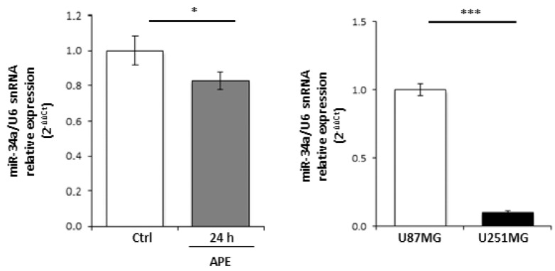
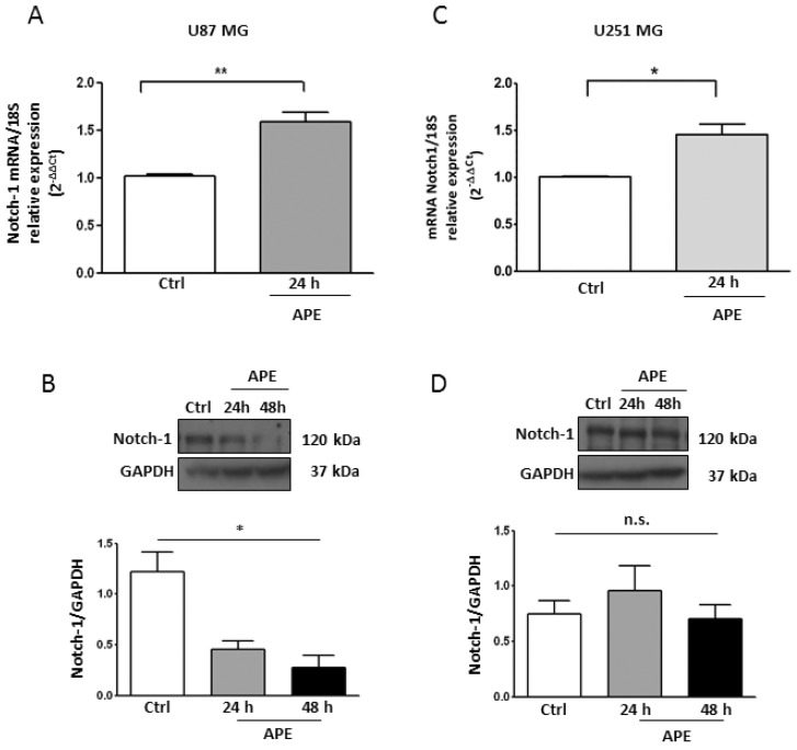
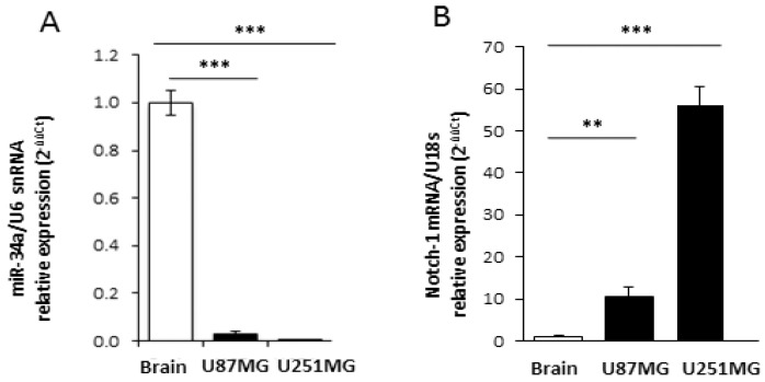
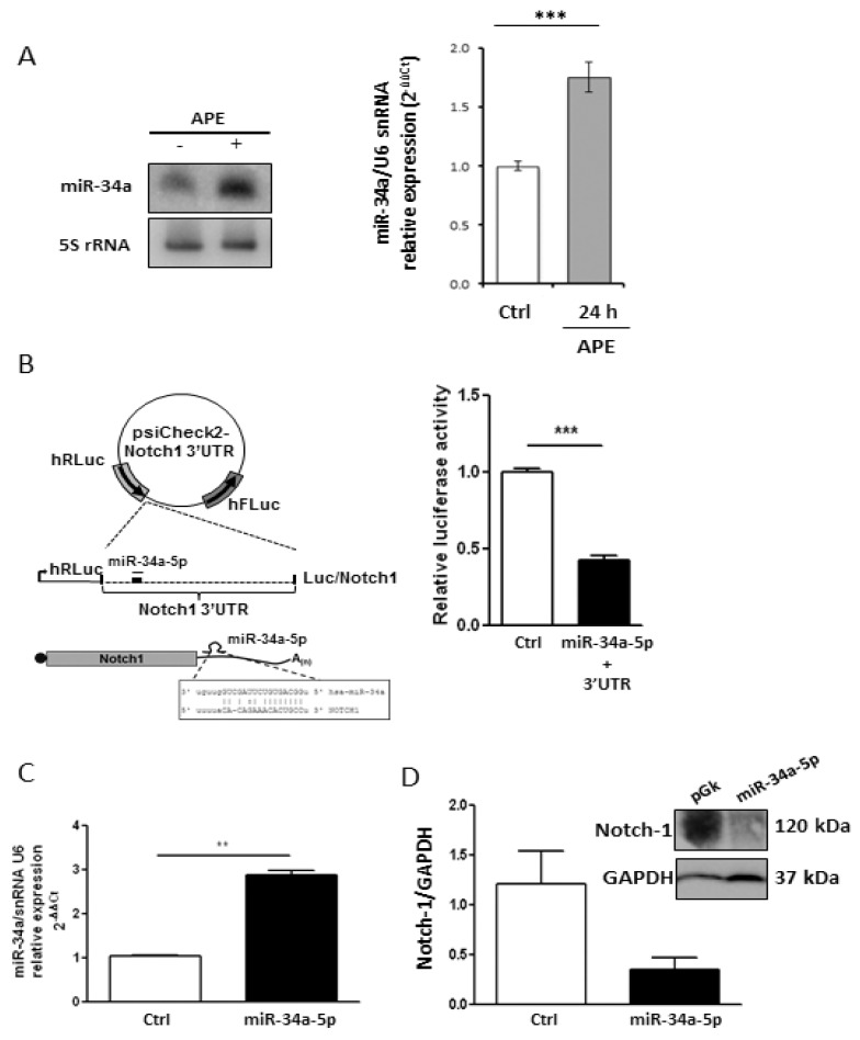
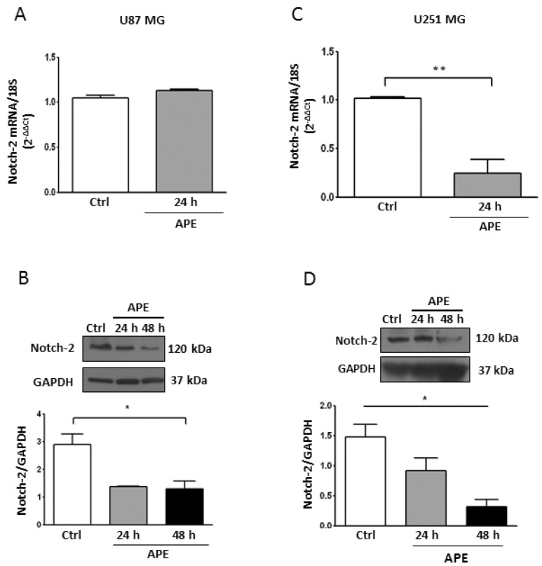
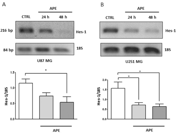
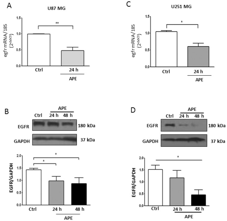
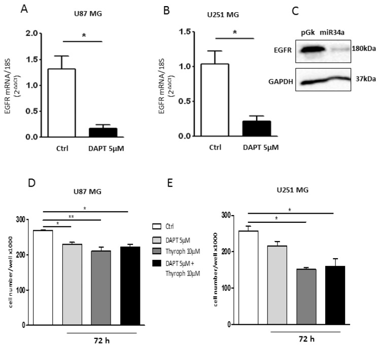
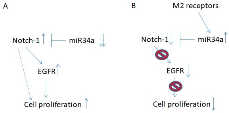
Glioblastoma (GBM) is the most aggressive human brain tumor. The high growth potential and decreased susceptibility to apoptosis of the glioma cells is mainly dependent on genetic amplifications or mutations of oncogenic or pro-apoptotic genes, respectively. We have previously shown that the activation of the M2 acetylcholine muscarinic receptors inhibited cell proliferation and induced apoptosis in two GBM cell lines and cancer stem cells. The aim of this study was to delve into the molecular mechanisms underlying the M2-mediated cell proliferation arrest. Exploiting U87MG and U251MG cell lines as model systems, we evaluated the ability of M2 receptors to interfere with Notch-1 and EGFR pathways, whose activation promotes GBM proliferation. We demonstrated that the activation of M2 receptors, by agonist treatment, counteracted Notch and EGFR signaling, through different regulatory cascades depending, at least in part, on p53 status. Only in U87MG cells, which mimic p53-wild type GBMs, did M2 activation trigger a molecular circuitry involving p53, Notch-1, and the tumor suppressor mir-34a-5p. This regulatory module negatively controls Notch-1, which affects cell proliferation mainly through the Notch-1/EGFR axis. Our data highlighted, for the first time, a molecular circuitry that is deregulated in the p53 wild type GBM, based on the cross-talk between M2 receptor and the Notch-1/EGFR pathways, mediated by mir-34a-5p.

Keywords: EGFR; M2 muscarinic receptors; Notch-1; glioblastoma; mir-34a-5p; p53.


Conflict of interest statement

The authors declare no conflict of interests.

Figures

Figure A1
Figure A1
(A) Expression of miR-34-5p in GBM cell lines upon APE treatment. qRT-PCR (real time PCR analysis) expression analysis of miR-34-5p in U251MG untreated or treated for 24 h with 100 μM APE. (B) Relative expression of miR-34a-5p in U251MG cells versus U87MG cells. snRNA U6 was used as the internal standard (* p < 0.05; *** p < 0.001; t-test).
Figure 1
Figure 1
Notch-1 Expression in GBM cell lines. Real time RT-PCR and Western blot analysis (A and B, respectively) for Notch-1 in U87MG and in U251MG cells (C and D, respectively) cultured in the absence or presence of 100 μM APE for 24 and 48 h. Representative blots are shown from three independent experiments. GAPDH was used as the internal reference protein (* p < 0.05, ** p < 0.01).
Figure 2
Figure 2
Expression of Notch-1 and miR-34a-5p in GBM cell lines and human brain. Real time RT-PCR analysis of miR-34a-5p (A) and Notch-1 (B) relative expression in U87MG or U251MG cell lines (black bars) compared to human normal brain (white bar). snRNA U6 and 18S were respectively used as the internal standard (** p < 0.01; ***p < 0.001; t-test).
Figure 3
Figure 3
Analysis of Notch-1/miR-34-5p interaction. (A) Analysis of miR-34a-5p expression in U87MG cells, treated with 100 μM APE, by Northern blot (left) and real time RT-PCR (right) (*** p < 0.001 t-test); (B) Upper scheme: representation of Luc/Notch reporter construct. MiR is indicated as a thin line, miRNA response element as a thick line. Lower scheme: representation of 3′UTR region (with related sequences) binding mir-34a-5p. Right panel: luciferase activity (Renilla/Firefly ratio) of Notch-1 3′UTR reporter gene in HEK293 cells transfected for 48 h with the mir-34a-5p expressing vector or with empty vector used as control (Ctrl). Data are presented as mean ± SD from at least three different experiments. (*** p < 0.001 t-test); (C) mir-34a-5p over-expression after transfection in U87 cells (** p < 0.01 t-test); (D) Western blot analysis for Notch-1 levels in U87MG cells transfected with mir-34a-5p.
Figure 4
Figure 4
Notch-2 Expression in GBM cell lines. Real time RT-PCR and Western blot analysis (A,B, respectively) of Notch-2 in U87MG. Parallel analyses were performed in U251MG cells (C,D, respectively). Both lines were untreated or treated with 100 μM APE for 24 and 48 h. Representative blots are shown from three independent experiments. GAPDH was used as the internal reference protein. (* p < 0.05 One-way ANOVA test, ** p < 0.01 t-test).
Figure 5
Figure 5
Hes-1 Expression in GBM cell lines. RT-PCR analysis of Hes-1 in U87MG (A) and U251MG (B) cells treated with 100 μM APE for 24 and 48 h. The graphs show the densitometric analysis of the bands normalized for the housekeeping 18S. The OD is the mean ± SEM of three independent experiments. (* p < 0.05; One-way ANOVA test).
Figure 6
Figure 6
EGFR Expression in GBM cell lines. Real time RT-PCR and Western blot analysis (A,B, respectively) of EGFR in U87MG. Parallel analyses were performed in U251MG cells (C,D, respectively). Both lines were untreated or treated with 100 μM APE for 24 and 48 h. Representative blots are shown from three independent experiments. GAPDH was used as the internal reference protein. (* p < 0.05; ** p < 0.01; t-test and one-way ANOVA test).
Figure 7
Figure 7
Effect of Notch/EGFR inhibition on GBM cell growth. Real time RT-PCR analysis of EGFR expression in U87MG (A) and U251MG (B) cells treated with 5 µM DAPT for 24 h; (C) Western blot analysis for EGFR in U87MG cells transfected with mir-34a-5p. GAPDH was used as the internal reference protein (* p < 0.05; t-test); (D,E). Analysis of the cell growth in U87MG and U251MG cells, respectively, treated with 5 µM DAPT, or 10 µM Tyrph or 5 µM DAPT plus 10 µM Tyrph (* p < 0.05; ** p < 0.01; t-test and one-way ANOVA test).
Figure 8
Figure 8
Schematic representation of the Notch-1/EGFR axis in U87 cells. (A) In U87 cells, the downregulated expression of mir-34a-5p caused the up-regulation of Notch-1 expression with consequent increased expression of EGFR. The up-regulation of these two receptors causes increased cell proliferation. (B) M2 muscarinic receptors up-regulate the expression of mir-34a-5p, which prevents the increased levels of Notch-1. The down-regulation of Notch-1 negatively affects cell proliferation by the down-regulated expression of EGFR.

References

    1. Kleihues P., Louis D.N., Scheithauer B.W., Rorke L.B., Reifenberger G., Burger P.C., Cavenee W.K. The WHO classification of tumors of the nervous system. J. Neuropathol. Exp. Neurol. 2002;61:215–225. doi: 10.1093/jnen/61.3.215. - DOI - PubMed
    1. Mao H., Lebrun D.G., Yang J., Zhu V.F., Li M. Deregulated signaling pathways in glioblastoma multiforme: Molecular mechanisms and therapeutic targets. Cancer Investig. 2012;30:48–56. doi: 10.3109/07357907.2011.630050. - DOI - PMC - PubMed
    1. Yuan X., Wu H., Xu H., Xiong H., Chu Q., Yu S., Wu G.S., Wu K. Notch signaling: An emerging therapeutic target for cancer treatment. Cancer Lett. 2015;369:20–27. doi: 10.1016/j.canlet.2015.07.048. - DOI - PubMed
    1. Jarriault S., Brou C., Logeat F., Schroeter E.H., Kopan R., Israel A. Signalling downstream of activated mammalian Notch. Nature. 1995;377:355–358. doi: 10.1038/377355a0. - DOI - PubMed
    1. Yahyanejad S., King H., Iglesias V.S., Granton P.V., Barbeau L.M.O., van Hoof S.J., Groot A.J., Habets R., Prickaerts J., Chalmers A.J., et al. NOTCH blockade combined with radiation therapy and temozolomide prolongs survival of orthotopic glioblastoma. Oncotarget. 2016;7:41251–41264. doi: 10.18632/oncotarget.9275. - DOI - PMC - PubMed

LinkOut - more resources