. 2018 May 31;19(6):1638.
doi: 10.3390/ijms19061638.

Anti-Apoptosis and Anti-Fibrosis Effects of Eriobotrya Japonica in Spontaneously Hypertensive Rat Hearts

Affiliations

Anti-Apoptosis and Anti-Fibrosis Effects of Eriobotrya Japonica in Spontaneously Hypertensive Rat Hearts

Jui-Ting Chiang et al. Int J Mol Sci. .

Abstract

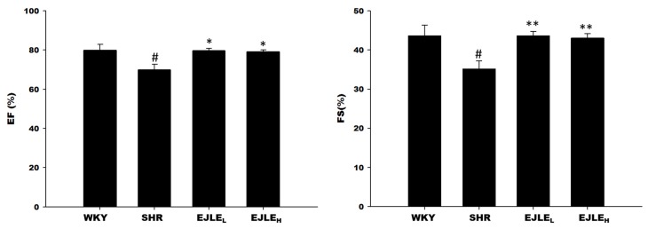
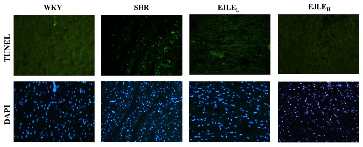
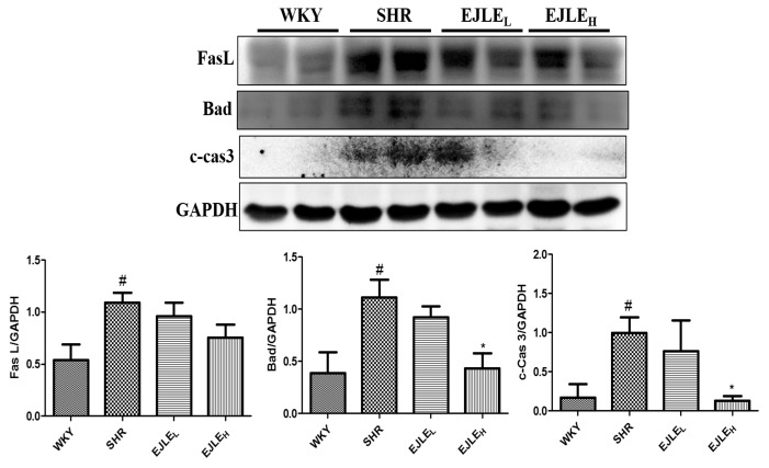
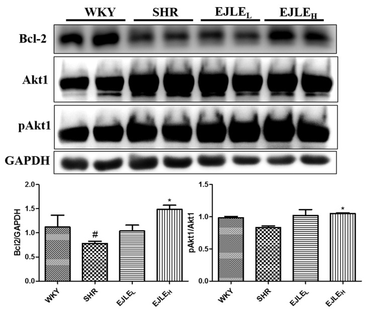
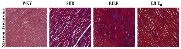
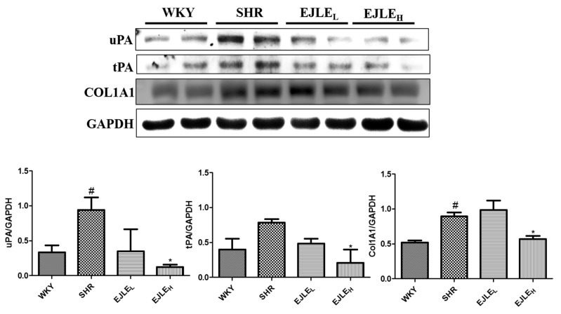
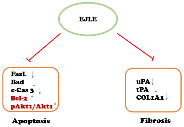
Myocardial apoptosis and fibrosis represent important contributing factors for development of hypertension-induced heart failure. The present study aims to investigate the potential effects of Eriobotrya japonica leaf extract (EJLE) against hypertension-induced cardiac apoptosis and fibrosis in spontaneously hypertensive rats (SHRs). Twelve-week-old male rats were randomly divided into four different groups; control Wistar Kyoto (WKY) rats, hypertensive SHR rats, SHR rats treated with a low dose (100 mg/kg body weight) of EJLE and SHR rats treated with a high dose (300 mg/kg body weight) of EJLE. Animals were acclimatized for 4 weeks and thereafter were gastric fed for 8 weeks with two doses of EJLE per week. The rats were then euthanized following cardiac functional analysis by echocardiography. The cardiac tissue sections were examined by Terminal Deoxynucleotidyl Transferase-Mediated Deoxyuridine Triphosphate (dUTP) Nick End-Labeling (TUNEL) assay, histological staining and Western blotting to assess the cardio-protective effects of EJ in SHR animals. Echocardiographic measurements provided convincing evidence to support the ability of EJ to ameliorate crucial cardiac functional characteristics. Furthermore, our results reveal that supplementation of EJLE effectively attenuated cardiac apoptosis and fibrosis and also enhanced cell survival in hypertensive SHR hearts. Thus, the present study concludes that EJLE potentially provides cardio-protective effects against hypertension-induced cardiac apoptosis and fibrosis in SHR animals.

Keywords: Eriobotrya japonica; SHRs; apoptosis; fibrosis.


Conflict of interest statement

The authors declare no conflict of interest.

Figures

Figure 1
Figure 1
Effect of EJLE on cardiac functional characteristics of SHR animals according to echocardiographic assessment. Differences in Ejection Fraction (EF) and Fraction shortening (FS) levels determined by echocardiography in normotensive Wistar Kyoto rats (WKY), spontaneously hypertensive rats (SHRs) and SHRs supplemented with low dose (EJLEL) and high dose (EJLEH). The values are the means ± S.D. All measurements were performed post EJLE treatment. # (p < 0.05) indicate significant differences when compared to normotensive WKY group (SHRs vs. WKY); whereas * (p < 0.05) and ** (p < 0.01) indicate significant differences when compared to SHR group.
Figure 2
Figure 2
Effect of EJLE on cardiac apoptosis in SHR animals. Representative photomicrographs of TUNEL and DAPI stained nuclei in heart tissue sections of normotensive (WKY), spontaneously hypertensive rats (SHRs) and SHRs supplemented with low dose (EJLEL) and high dose (EJLEH) of EJLE. The images were acquired at 400× magnification.
Figure 3
Figure 3
Effect of EJLE on apoptotic signaling mediators in the heart tissue sections of SHR animals. Representative Western blot depicting the changes in the levels of FasL, Bad, c-Cas3 proteins involved in apoptosis in normotensive rats (WKY), spontaneously hypertensive rats (SHRs) and SHRs supplemented with low dose (EJLEL) and high dose (EJLEH) of EJLE. Results were analyzed by one-way ANOVA using Tukey test with GraphPad Prism software (Version 5.0). # (p < 0.05) indicate significant differences when compared to normotensive WKY group (SHRs vs. WKY); whereas * (p < 0.05) indicate significant differences when compared to SHR group.
Figure 4
Figure 4
Effect of EJLE on the survival markers in the heart tissue sections of SHR animals. Representative Western blot depicting the changes in the levels of Bcl-2, Akt1, and pAkt1 proteins involved in the survival pathways in normotensive rats (WKY), spontaneously hypertensive rats (SHRs) and SHRs supplemented with low dose (EJLEL) and high dose (EJLEH). Results were analyzed by one-way ANOVA using Tukey test with GraphPad Prism software (Version 5.0). # (p < 0.05) indicate significant differences when compared to normotensive WKY group (SHR vs. WKY); whereas * (p < 0.05) indicate significant differences when compared to SHR group.
Figure 5
Figure 5
Effect of EJLE on fibrosis in the heart of SHR animals. Representative Masson’s trichrome photomicrograph depicting the effect of EJLE on the heart tissue sections of normotensive rats (WKY), spontaneously hypertensive rats (SHRs) and SHRs supplemented with low dose (EJLEL) and high dose (EJLEH) of EJLE. The heart tissue sections from all animals were processed for Masson’s trichrome staining as detailed in the materials and methods and images were acquired through microscope at 400× magnification.
Figure 6
Figure 6
Effect of EJLE on the fibrosis associated proteins in the heart tissue section of SHR animals. Representative Western blot depicting the changes in the levels of COL1A1, tPA, and uPA proteins involved in fibrosis in normotensive rats (WKY), spontaneously hypertensive rats (SHRs) and SHRs supplemented with low dose (EJLEL) and high dose (EJLEH) of EJLE. Results were analyzed by one-way ANOVA using Tukey test with GraphPad Prism software (Version 5.0). # (p < 0.05) indicate significant differences when compared to normotensive WKY group (SHR vs. WKY); whereas * (p < 0.05) indicate significant differences when compared to SHR group.
Figure 7
Figure 7
Schematic outline representing the effect of EJLE on cardiac apoptosis mediators (FasL, Bad, c-Cas 3, and anti-apoptotic/survival markers Bcl2 and Akt1) and fibrosis mediators (uPA, tPA, COL1A1) in model animals. Arrow represents proteins modulated by EJLE. The T bar represents attenuation of cardiac apoptosis and fibrosis through supplementation of EJLE, whereas the upward and downward arrows represent the protein expressions pattern.

References

    1. Bromfield S., Muntner P. High blood pressure: The leading global burden of disease risk factor and the need for worldwide prevention programs. Curr. Hypertens. Rep. 2013;15:134–136. - PMC - PubMed
    1. Diez J., Fortuno M.A., Ravassa S. Apoptosis in hypertensive cardiopathy. Revista Espanola de Cardiologia. 1999;52:18–24. - PubMed
    1. Huang C.Y., Kuo W.W., Yeh Y.L., Ho T.J., Lin J.Y., Lin D.Y., Chu C.H., Tsai F.J., Tsai C.H., Huang C.Y. ANG II promotes IGF-IIR expression and cardiomyocyte apoptosis by inhibiting HSF1 via JNK activation and SIRT1 degradation. Cell Death Differ. 2014;21:1262–1274. - PMC - PubMed
    1. Chu C.H., Lo J.F., Hu W.S., Lu R.B., Chang M.H., Tsai F.J., Tsai C.H., Weng Y.S., Tzang B.S., Huang C.Y. Histone acetylation is essential for ANG-II-induced IGF-IIR gene expression in H9c2 cardiomyoblast cells and pathologically hypertensive rat heart. J. Cell Physiol. 2012;227:259–268. doi: 10.1002/jcp.22728. - DOI - PubMed
    1. Lee S.D., Chu C.H., Huang E.J., Lu M.C., Liu J.Y., Liu C.J., Hsu H.H., Lin J.A., Kuo W.W., Huang C.Y. Roles of insulin-like growth factor II in cardiomyoblast apoptosis and in hypertensive rat heart with abdominal aorta ligation. Am. J. Physiol. Endocrinol. Metab. 2006;291:E306–E314. doi: 10.1152/ajpendo.00127.2005. - DOI - PubMed

MeSH terms

LinkOut - more resources