Review
. 2018 May 31;7(6):51.
doi: 10.3390/cells7060051.

Fluorescent, Bioluminescent, and Optogenetic Approaches to Study Excitable Physiology in the Single Cardiomyocyte

Affiliations
Review

Fluorescent, Bioluminescent, and Optogenetic Approaches to Study Excitable Physiology in the Single Cardiomyocyte

Connor N Broyles et al. Cells. .

Abstract

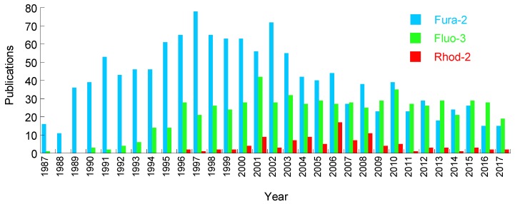
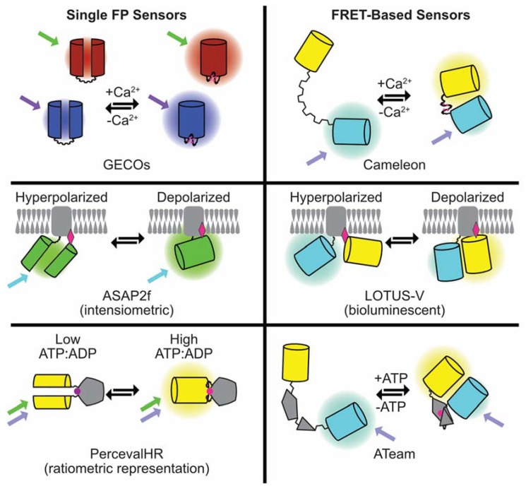
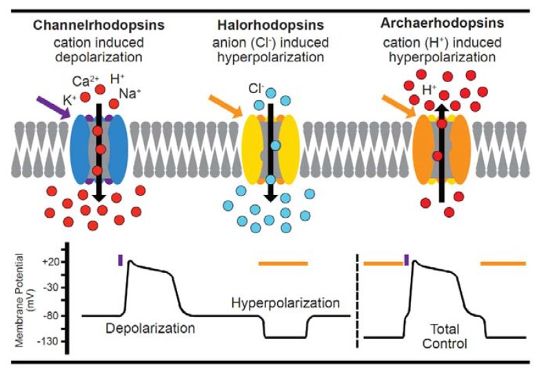
This review briefly summarizes the single cell application of classical chemical dyes used to visualize cardiomyocyte physiology and their undesirable toxicities which have the potential to confound experimental observations. We will discuss, in detail, the more recent iterative development of fluorescent and bioluminescent protein-based indicators and their emerging application to cardiomyocytes. We will discuss the integration of optical control strategies (optogenetics) to augment the standard imaging approach. This will be done in the context of potential applications, and barriers, of these technologies to disease modelling, drug toxicity, and drug discovery efforts at the single-cell scale.

Keywords: FRET; adult ventricular cardiomyocyte; bioluminescence; calcium; cardiomyocyte; chemical dye; excitable cell physiology; fluorescent protein; iPS-cardiomyocyte; optogenetics; voltage.


Conflict of interest statement

The authors declare no conflict of interest.

Figures

Figure 1
Figure 1
Annual publications imaging cardiomyocytes or heart tissue using the fluorescent Ca2+ indicators fura-2, fluo-4, and Rhod-2. Metrics extracted from CSV data at Pubmed.
Figure 2
Figure 2
Summary of the composition and mode of action of the genetically-encoded fluorescent sensors for Ca2+, voltage, and ATP discussed in the article.
Figure 3
Figure 3
A summary of optogenetic actuators. Depolarizing channelrhodopsins, e.g., ChR2, open in response to light in the blue-green spectrum allowing positively-charged ions into the cell, raising the membrane potential and triggering the depolarization threshold. Conversely, inhibitory channels like Halorhodopsin (which pumps negatively-charged ions in) or Archaerhodopsin (which pumps protons out) hyperpolarize excitable membranes. Inhibitory channel activity is controlled by light in the green-red spectrum. The lower panel stylizes the effect on cell membrane potential by blue or orange pulses of light to depolarize, or hyperpolarize the cell. The two approaches can be combined to hyperpolarize, and then activate, the cell [95]. A number of reporter strategies discussed in this review can be integrated with stimulatory optical control [42,60,68,69,78,81,83,85,97].

References

    1. Ridgway E.B., Ashley C.C. Calcium transients in single muscle fibers. Biochem. Biophys. Res. Commun. 1967;29:229–234. doi: 10.1016/0006-291X(67)90592-X. - DOI - PubMed
    1. Tsien R.Y. New calcium indicators and buffers with high selectivity against magnesium and protons: Design, synthesis, and properties of prototype structures. Biochemistry. 1980;19:2396–2404. doi: 10.1021/bi00552a018. - DOI - PubMed
    1. Tsien R., Pozzan T. Measurement of cytosolic free Ca2+ with quin2. Methods Enzymol. 1989;172:230–262. - PubMed
    1. Powell T., Tatham P.E., Twist V.W. Cytoplasmic free calcium measured by quin2 fluorescence in isolated ventricular myocytes at rest and during potassium-depolarization. Biochem. Biophys. Res. Commun. 1984;122:1012–1020. doi: 10.1016/0006-291X(84)91192-6. - DOI - PubMed
    1. Sheu S.S., Sharma V.K., Banerjee S.P. Measurement of cytosolic free calcium concentration in isolated rat ventricular myocytes with quin 2. Circ. Res. 1984;55:830–834. doi: 10.1161/01.RES.55.6.830. - DOI - PubMed

LinkOut - more resources