. 2018 Jun 1:11:253-262.
doi: 10.1016/j.omtn.2018.02.006. Epub 2018 Mar 30.

Time-Restricted PiggyBac DNA Transposition by Transposase Protein Delivery Using Lentivirus-Derived Nanoparticles

Affiliations

Time-Restricted PiggyBac DNA Transposition by Transposase Protein Delivery Using Lentivirus-Derived Nanoparticles

Kristian Alsbjerg Skipper et al. Mol Ther Nucleic Acids. .

Abstract

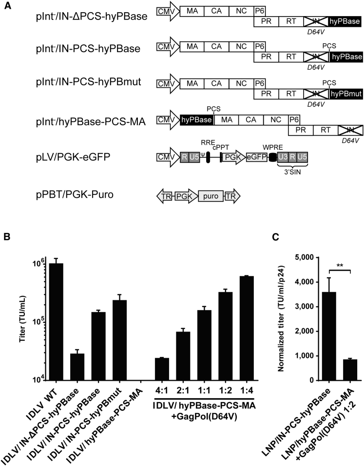
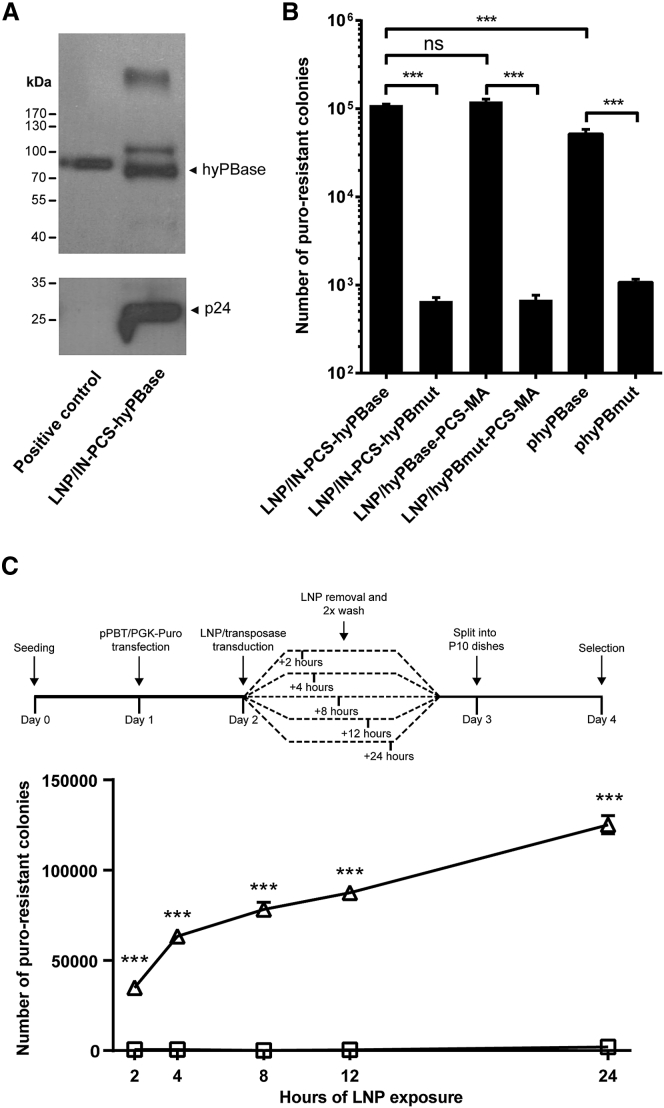
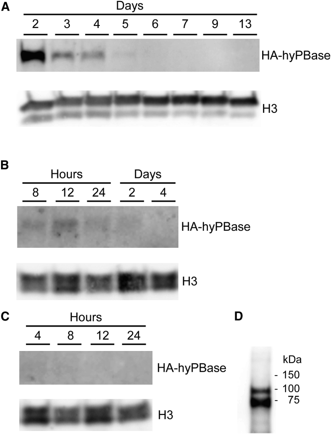
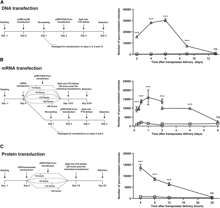
Continuous innovation of revolutionizing genome engineering technologies calls for an intensified focus on new delivery technologies that not only match the inventiveness of genome editors but also enable the combination of potent delivery and time-restricted action of genome-modifying bits and tools. We have previously demonstrated the use of lentivirus-derived nanoparticles (LNPs) as a protein delivery vehicle, incorporating and transferring DNA transposases, designer nucleases, or RNA-guided endonucleases fused to the N terminus of the Gag/GagPol polypeptide. Here, we establish LNP-directed transfer of the piggyBac DNA transposase protein by fusing the transposase to the integrase protein in the C-terminal end of GagPol. We show protein incorporation and proteolytic release of the DNA transposase within matured LNPs, resulting in high levels of DNA transposition activity in LNP-treated cells. Importantly, as opposed to conventional delivery methods based on transfection of plasmid DNA or in-vitro-transcribed mRNA, protein delivery by LNPs effectively results in time-restricted action of the protein (<24 hr) without compromising overall potency. Our findings refine LNP-directed piggyBac transposase delivery, at present the only available direct delivery strategy for this particular protein, and demonstrate a novel strategy for restricting and fine-tuning the exposure of the genome to DNA-modifying enzymes.

Keywords: DNA transposon; LNP; lentivirus; piggyBac; protein delivery; protein transduction.


Figures

Figure 1
Figure 1
Titer and Transfer Efficiency of IDLVs Packaged with Integrase-Fused hyPBase (A) Schematic representation of the constructs used in the study. (B) Evaluation of transduction titers of transposase-loaded IDLVs carrying vector RNA encoded by pLV/PGK-EGFP is shown. IDLVs with IN-fused PB transposase with or without the KARVL/AEAMS protease cleavage site (PCS) were packaged with a PGK-EGFP expression vector, and transduction titers were estimated by flow cytometry and compared to IDLVs packaged with MA-fused transposase in the presence of increasing amounts of wild-type Gag/GagPol (GagPol-D64V), as indicated by the ratios. A standard IDLV was furthermore included as a control. (C) Estimation of IDLV transfer efficiency is shown. The total amount of LNPs was estimated by p24 ELISA and used to normalize transduction titers quantified in (B) to estimate the relative transfer efficiency of IDLVs packaged with IN- or MA-fused PB transposase. cPPT, central polypurine tract; CA, capsid; IN, integrase; MA, matrix; NC, nucleocapsid; PR, protease; RT, reverse transcriptase; Ψ, psi packaging signal; RRE, rev response element; WPRE, woodchuck hepatitis virus post-transcriptional regulatory element. Data are presented as mean ± SEM and n ≥ 3.
Figure 2
Figure 2
Efficient and Rapid Cellular Uptake of hyPBase-Loaded LNPs (A) Evidence of LNP encapsidation of IN-fused hyPBase and subsequent proteolytic release of hyPBase. Western blot analysis on a LNP lysate incorporating an HA-tagged version of the hyPBase transposase is shown. Cell lysates from HEK293T cells transfected with 1 μg HA-hyPBase expressing plasmid served as positive control. (B) Colony formation after transposase delivery by hyPBase-loaded LNPs is shown. Initially, the PB transposon donor pPBT/PGK-Puro was delivered to HeLa cells by plasmid DNA transfection. The cells were subsequently treated with LNPs loaded with either IN- or MA-fused PB transposase, and transposase activity was estimated by quantification of puromycin-resistant colonies. (C) Limited exposure demonstrates rapid cellular uptake of LNPs. HeLa cells were exposed to PB-loaded LNPs for 2, 4, 8, 12, or 24 hr after delivery of the pPBT/PGK-Puro PB transposon donor. Transposase activity was subsequently estimated as described in (B). Triangles, LNP/hyPBase; squares, LNP/hyPBmut. Data are presented as mean ± SEM and n ≥ 3.
Figure 3
Figure 3
Western Blot Analysis of hyPBase Protein Levels over Time (A–C) HeLa cell lysates were analyzed by western blotting at the indicated time points after delivery of HA-tagged versions of the hyPBase transposase by either plasmid DNA transfection of 100 ng of a hyPBase expression vector (A), mRNA transfection of 150 ng in-vitro-transcribed hyPBase mRNA (B), or protein transduction of 400 ng P24 hyPBase-loaded LNPs (C). (D) A LNP lysate was furthermore included to verify the presence of hyPBase in the LNPs. For all blots, H3 was used as a loading control.
Figure 4
Figure 4
Determination of the Temporal Pattern of Protein Activity after Transposase Delivery by LNPs Compared to Standard Delivery Methods HeLa cells were transfected with 900 ng of the pPBT/PGK-Puro transposon donor, and subsequently, PB transposase was delivered to the cells by either plasmid DNA transfection (A), transfection of in-vitro-transcribed mRNA (B), or transduction of transposase-loaded LNPs (C) at the indicated time points. Detailed delivery schemes can be seen in the left panels of (A), (B), and (C). Transposase activity was subsequently estimated as described earlier. Triangles, hyPBase; squares, hyPBmut. Data are presented as mean ± SEM and n ≥ 3.

References

    1. De Ravin S.S., Reik A., Liu P.Q., Li L., Wu X., Su L., Raley C., Theobald N., Choi U., Song A.H. Targeted gene addition in human CD34(+) hematopoietic cells for correction of X-linked chronic granulomatous disease. Nat. Biotechnol. 2016;34:424–429. - PMC - PubMed
    1. Dever D.P., Bak R.O., Reinisch A., Camarena J., Washington G., Nicolas C.E., Pavel-Dinu M., Saxena N., Wilkens A.B., Mantri S. CRISPR/Cas9 β-globin gene targeting in human haematopoietic stem cells. Nature. 2016;539:384–389. - PMC - PubMed
    1. Skipper K.A., Mikkelsen J.G. Delivering the goods for genome engineering and editing. Hum. Gene Ther. 2015;26:486–497. - PubMed
    1. Cyranoski D. Chinese scientists to pioneer first human CRISPR trial. Nature. 2016;535:476–477. - PubMed
    1. Tebas P., Stein D., Tang W.W., Frank I., Wang S.Q., Lee G., Spratt S.K., Surosky R.T., Giedlin M.A., Nichol G. Gene editing of CCR5 in autologous CD4 T cells of persons infected with HIV. N. Engl. J. Med. 2014;370:901–910. - PMC - PubMed