. 2018 Jun 1:11:429-440.
doi: 10.1016/j.omtn.2018.03.007. Epub 2018 Mar 16.

CRISPR/Cas9 Mediated Disruption of the Swedish APP Allele as a Therapeutic Approach for Early-Onset Alzheimer's Disease

Affiliations

CRISPR/Cas9 Mediated Disruption of the Swedish APP Allele as a Therapeutic Approach for Early-Onset Alzheimer's Disease

Bence György et al. Mol Ther Nucleic Acids. .

Abstract

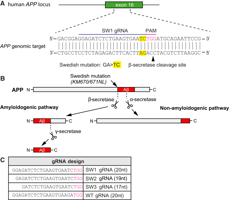
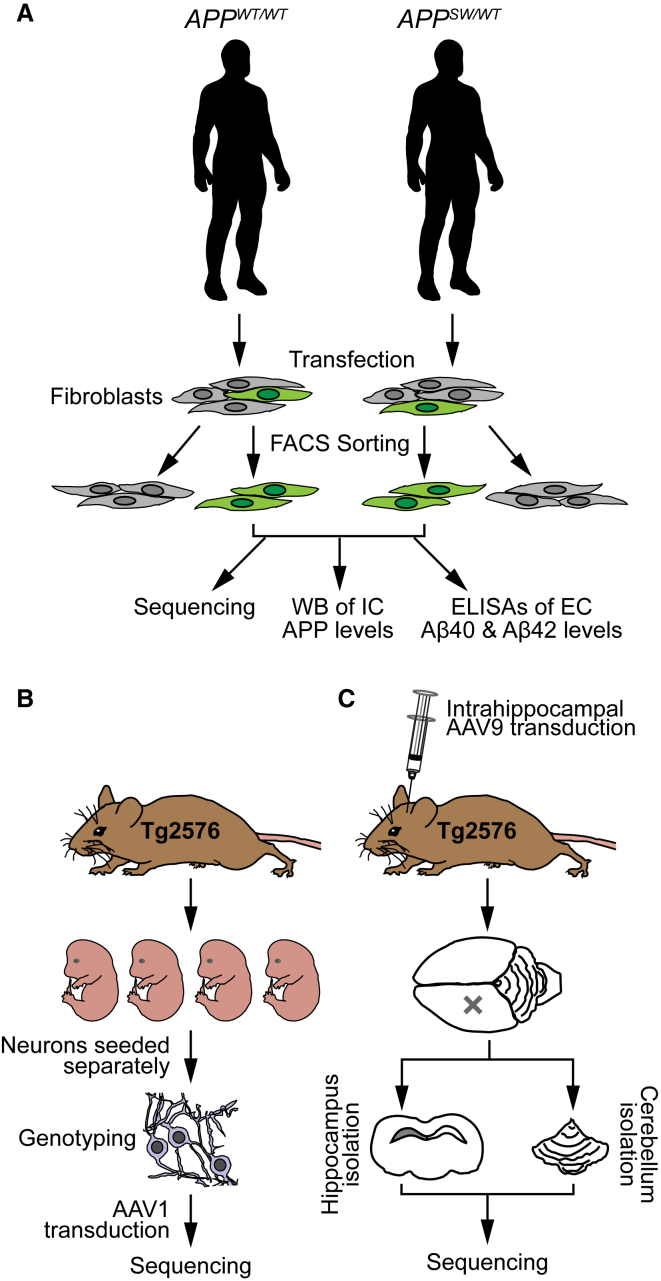
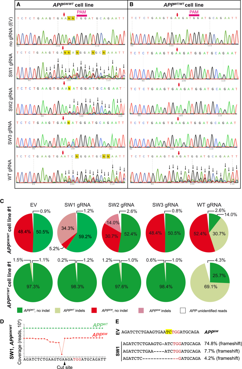
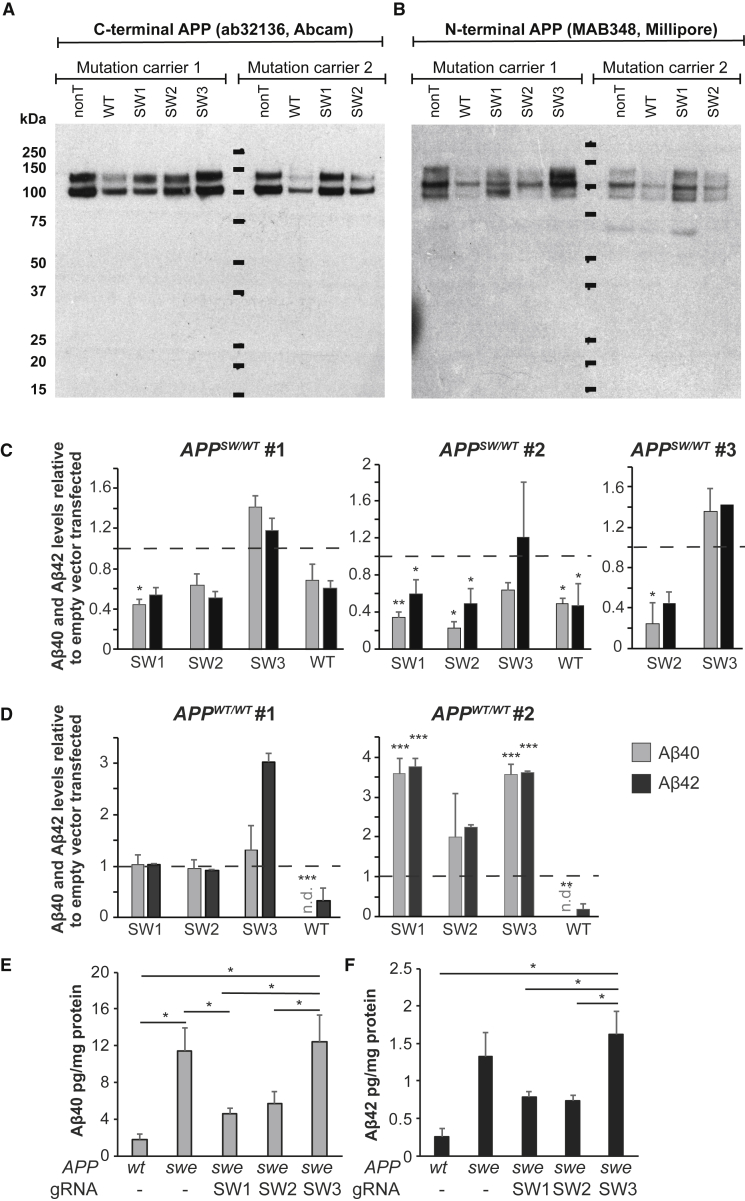
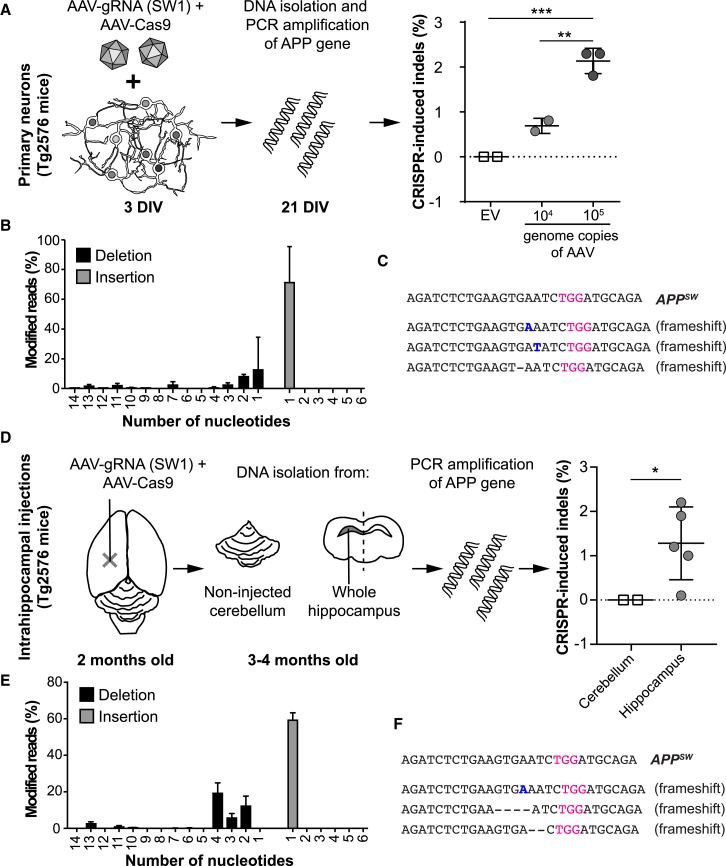
The APPswe (Swedish) mutation in the amyloid precursor protein (APP) gene causes dominantly inherited Alzheimer's disease (AD) as a result of increased β-secretase cleavage of the amyloid-β (Aβ) precursor protein. This leads to abnormally high Aβ levels, not only in brain but also in peripheral tissues of mutation carriers. Here, we selectively disrupted the human mutant APPSW allele using CRISPR. By applying CRISPR/Cas9 from Streptococcus pyogenes, we generated allele-specific deletions of either APPSW or APPWT. As measured by ELISA, conditioned media of targeted patient-derived fibroblasts displayed an approximate 60% reduction in secreted Aβ. Next, coding sequences for the APPSW-specific guide RNA (gRNA) and Cas9 were packaged into separate adeno-associated viral (AAV) vectors. Site-specific indel formation was achieved both in primary neurons isolated from APPSW transgenic mouse embryos (Tg2576) and after co-injection of these vectors into hippocampus of adult mice. Taken together, we here present proof-of-concept data that CRISPR/Cas9 can selectively disrupt the APPSW allele both ex vivo and in vivo-and thereby decrease pathogenic Aβ. Hence, this system may have the potential to be developed as a tool for gene therapy against AD caused by APPswe and other point mutations associated with increased Aβ.

Keywords: Alzheimer's disease; CRISPR; Swedish mutation; adeno-associated virus; amyloid precursor protein; amyloid-β; genome editing.


Figures

Figure 1
Figure 1
CRISPR Targeting of Swedish KM670/671NL APP and Its Effect on Aβ Generation (A) The Swedish KM670/671NL APP (APPswe) double-base change (yellow), causing familial Alzheimer’s disease (AD), is located near a potential Streptococcus pyogenes Cas9 PAM (NGG) site (purple). The mutation and gRNA target site is upstream of the β-secretase site. (B) In the amyloidogenic pathway, amyloid-β (Aβ) is produced via sequential cleavages of the amyloid precursor protein (APP) by β- and γ-secretases. No Aβ is generated upon α-secretase APP cleavage in the non-amyloidogenic pathway. The APPswe mutation is a better β-secretase substrate than the corresponding wild-type site, and individuals with this mutation develop AD as a consequence of elevated Aβ levels. (C) Three different gRNAs targeting the KM670/671NL site (SW1, SW2, and SW3) and one gRNA recognizing the wild-type sequence (WT) were evaluated. The PAM site is depicted in purple.
Figure 2
Figure 2
Overview of the Study Design (A) Fibroblasts were collected from human APPswe carriers and their non-affected relatives. The cells were transfected with S. pyogenes Cas9-2A-GFP and gRNAs. Successfully transfected cells were identified by GFP expression and sorted by FACS. Next, these cells were expanded in culture and analyzed by sequencing, western blot (WB) (for intracellular [IC] levels of APP), and ELISA (for extracellular [EC] secretion of Aβ40 and Aβ42). (B) Embryos from time-pregnant Tg2576 APPswe transgenic mice (embryonic day 14 [E14]–17) were used to generate primary cortical neuronal culture. The transgenic cultures were co-transduced for one day with AAV1-Cas9 and either AAV1-gRNA(SW1) at 3 days in vitro (DIV). At 21 DIV, the cells were collected for sequencing. (C) Adult Tg2576 transgenic mice were co-transduced unilaterally in hippocampus with AAV9-Cas9 and AAV9-gRNA(SW1). After 1 or 2 months, the mice were sacrificed and the injected hippocampi and non-injected cerebelli (as controls) were isolated for genomic DNA sequencing.
Figure 3
Figure 3
CRISPR/Cas9 Mediates Disruption of APPSW or APPWT Allele in Patient-Derived Fibroblasts (A and B) Sanger sequencing of the APP gene from mutation carrier No. 1 and control line No. 1 transfected with Cas9-2A-GFP and gRNA targeting the APPSW (SW1, SW2, and SW3) or APPWT allele (WT), respectively. GFP-expressing cells were sorted 3 days after transfection. On the APPSW/WT cell line (A), SW1, SW2, and WT gRNA were disruptive and created indels (as indicated by background peaks [i.e., heterogeneity in reads] downstream of the cut site, black arrows). On the APPWT/WT cell line (B), WT gRNA led to indel formation, whereas SW1, SW2, or SW3 gRNA did not. PAM, protospacer-adjacent motif. The purple arrow shows the predicted CRISPR cut site. (C) Targeted deep sequencing of the APP allele from CRISPR-treated APPSW/WT fibroblasts (top pie charts) and control APPWT/WT fibroblasts (bottom pie charts) is shown. Colors indicate mutant (red), wild-type (green), mutant with indels (light red), and wild-type with indels (light green) reads. For some reads, we were not able to determine their origins as mutant or wild-type (due to sequencing errors or larger deletions), and these are marked as white areas on the pie charts. SW1 and SW2 gRNA resulted in indel formation in the APPSW, but not in the APPWT allele. WT gRNA resulted in indel formation only in the APPWT allele in APPSW/WT cells and APPWT/WT cells. SW3 did not lead to indel formation in any of the alleles. (D) Coverage of the APP allele in APPSW/WT cells, treated with Cas9 and SW1 gRNA, is shown. Deletions were detectable adjacent to the CRISPR cut site on the APPSW allele (red trace). No deletions were present on the APPWT allele. The PAM site is marked in red. (E) The most frequent reads in the APPSW/WT fibroblasts treated with Cas9 and SW1 gRNA are shown. PAM sites are marked with red, and the APPswe mutation is marked in yellow. The percentages refer to the fraction of reads that contained any indels.
Figure 4
Figure 4
CRISPR/Cas9 against APPSW and APPWT Reduces the Levels of Aβ (A and B) Western blot of C terminus APP (C-APP) (A) and N terminus APP (N-APP) (B) of lysates from two mutation carriers. For quantification, see Figure S1. (C and D) Levels of secreted Aβ40 and Aβ42 in conditioned media of human CRISPR/Cas9-treated and control fibroblasts, as measured by ELISA. (C) shows APPSW/WT fibroblasts and (D) APPWT/WT fibroblasts. Data are normalized to total protein content and plotted as a relative value compared to empty vector transfected cells (value of empty vector [ = 1] is represented by dashed lines, mean ± SEM). The numbers (No. 1, No. 2, and No. 3) above the charts show cell lines from different patients or non-affected individuals. SW1, SW2, and SW3 are different gRNAs against the APPSW allele, whereas WT is a gRNA against the APPWT allele. n.d., not determined (below detection limit); *p < 0.05; **p < 0.01; ***p < 0.001. (E and F) Average of absolute levels (mean ±SEM) of Aβ40 (E) and Aβ42 (F) as measured by ELISA. swe, mutant lines; wt, wild-type lines; *p < 0.05 (one-way ANOVA with Tukey’s post hoc test).
Figure 5
Figure 5
CRISPR/Cas9 Can Specifically Target the APPSW Allele in Primary Neurons and in the Brain of Tg2576 Mice (A) Primary cortical neurons from Tg2576 mice (embryonic day 14–17) were co-transduced with exo-AAV1-Cas9 and exo-AAV1-gRNA (SW1) or exo-AAV1-Cas9 with exo-AAV-gRNA (empty vector [EV]) at 3 days in vitro (DIV). At 21 DIV, total DNA was collected and analyzed for indel formation by targeted high-throughput sequencing. The SW1-gRNA-treated cells showed a 0.7% and 2.3% indel formation using 104 and 105 genomic copies of AAV per cell, respectively (mean ± SD). In contrast, EV-treated cells did not display indels (**p < 0.01; ***p < 0.001; one-way ANOVA with post hoc Tukey’s test). (B) Indel profile in vitro (mean ± SEM). (C) The most abundant CRISPR-induced changes in vitro on cortical neurons. Of all the indels presented after treatment with SW1, 70.9% were a single nucleotide insertion (inserted nucleotide is depicted in blue), whereas only about 12.6% were a single nucleotide deletion The target PAM site is marked in purple; inserted nucleotides are marked in blue. (D) Two-month-old Tg2576 mice were injected unilaterally into hippocampus with exo-AAV9-Cas9 and exo-AAV9-gRNA (SW1), 4–8 weeks post-injection, DNA was collected and analyzed from total hippocampus and from cerebellum as control. An average indel percent of 1.3% was calculated after high-throughput sequencing compared to no indels identified in cerebellum (*p < 0.05; Mann-Whitney test). Mean ± SD is plotted. (E) Indel profile in vivo (mean ± SD). (F) The most abundant CRISPR-induced changes in vivo. The most common indel was a single-nucleotide insertion (60.3%), the second most common indel a five-nucleotide deletion (21.5%), and the third most common a double deletion (10.1%).

References

    1. Bertram L., McQueen M.B., Mullin K., Blacker D., Tanzi R.E. Systematic meta-analyses of Alzheimer disease genetic association studies: the AlzGene database. Nat. Genet. 2007;39:17–23. - PubMed
    1. Bertram L., Tanzi R.E. The genetics of Alzheimer’s disease. Prog. Mol. Biol. Transl. Sci. 2012;107:79–100. - PubMed
    1. Mullan M., Crawford F., Axelman K., Houlden H., Lilius L., Winblad B., Lannfelt L. A pathogenic mutation for probable Alzheimer’s disease in the APP gene at the N-terminus of beta-amyloid. Nat. Genet. 1992;1:345–347. - PubMed
    1. Citron M., Vigo-Pelfrey C., Teplow D.B., Miller C., Schenk D., Johnston J., Winblad B., Venizelos N., Lannfelt L., Selkoe D.J. Excessive production of amyloid beta-protein by peripheral cells of symptomatic and presymptomatic patients carrying the Swedish familial Alzheimer disease mutation. Proc. Natl. Acad. Sci. USA. 1994;91:11993–11997. - PMC - PubMed
    1. Johnston J.A., Cowburn R.F., Norgren S., Wiehager B., Venizelos N., Winblad B., Vigo-Pelfrey C., Schenk D., Lannfelt L., O’Neill C. Increased beta-amyloid release and levels of amyloid precursor protein (APP) in fibroblast cell lines from family members with the Swedish Alzheimer’s disease APP670/671 mutation. FEBS Lett. 1994;354:274–278. - PubMed