. 2018 Jun 1:11:475-484.
doi: 10.1016/j.omtn.2018.03.013. Epub 2018 Mar 30.

De Novo Synthesis of Elastin by Exogenous Delivery of Synthetic Modified mRNA into Skin and Elastin-Deficient Cells

Affiliations

De Novo Synthesis of Elastin by Exogenous Delivery of Synthetic Modified mRNA into Skin and Elastin-Deficient Cells

Mario Lescan et al. Mol Ther Nucleic Acids. .

Abstract

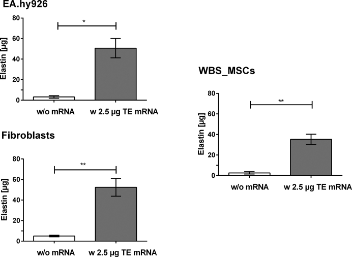
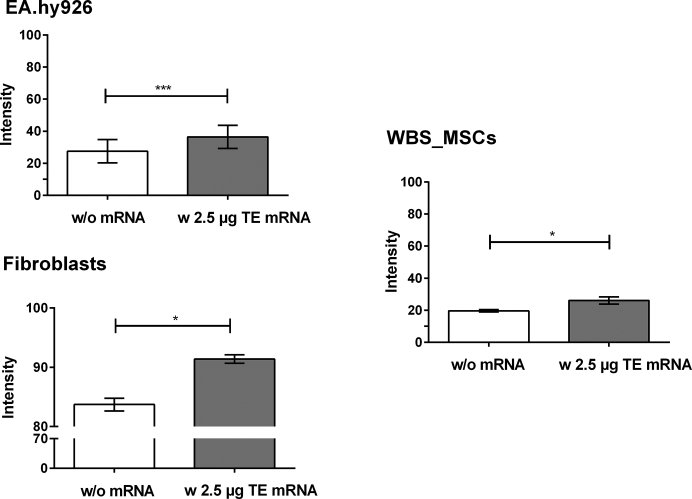
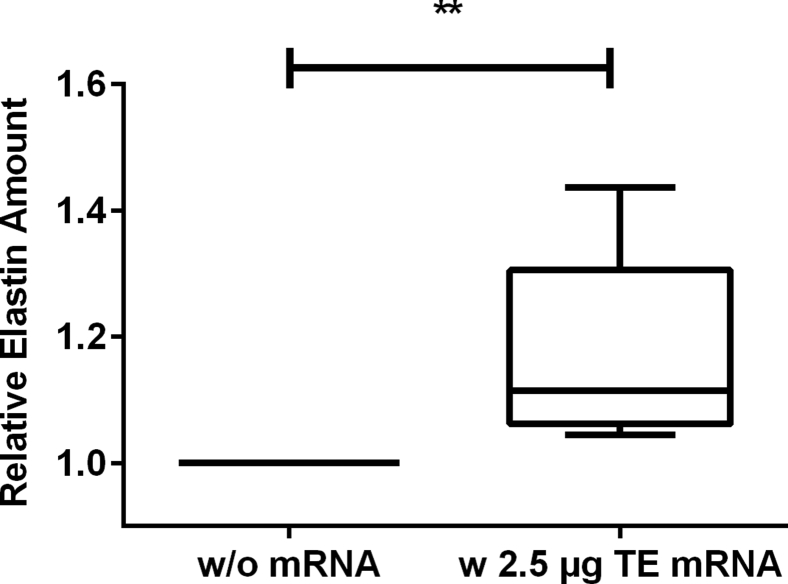
Elastin is one of the most important and abundant extracellular matrix (ECM) proteins that provide elasticity and resilience to tissues and organs, including vascular walls, ligaments, skin, and lung. Besides hereditary diseases, such as Williams-Beuren syndrome (WBS), which results in reduced elastin synthesis, injuries, aging, or acquired diseases can lead to the degradation of existing elastin fibers. Thus, the de novo synthesis of elastin is required in several medical conditions to restore the elasticity of affected tissues. Here, we applied synthetic modified mRNA encoding tropoelastin (TE) for the de novo synthesis of elastin and determined the mRNA-mediated elastin synthesis in cells, as well as ex vivo in porcine skin. EA.hy926 cells, human fibroblasts, and mesenchymal stem cells (MSCs) isolated from a patient with WBS were transfected with 2.5 μg TE mRNA. After 24 hr, the production of elastin was analyzed by Fastin assay and dot blot analyses. Compared with untreated cells, significantly enhanced elastin amounts were detected in TE mRNA transfected cells. The delivered synthetic TE mRNA was even able to significantly increase the elastin production in elastin-deficient MSCs. In porcine skin, approximately 20% higher elastin amount was detected after the intradermal delivery of synthetic mRNA by microinjection. In this study, we demonstrated the successful applicability of synthetic TE encoding mRNA to produce elastin in elastin-deficient cells as well as in skin. Thus, this auspicious mRNA-based integration-free method has a huge potential in the field of regenerative medicine to induce de novo elastin synthesis, e.g., in skin, blood vessels, or alveoli.

Keywords: Williams-Beuren syndrome; elastin; intradermal delivery; skin; synthetic mRNA.


Figures

Figure 1
Figure 1
Analysis of the Generated Synthetic TE mRNA and Cy3-Labeled TE mRNA by 1% Agarose Gel Electrophoresis (Lane 1) RNA marker, 400 ng of (lane 2) Cy3-labeled TE mRNA, (lane 3) modified, or (lane 4) unmodified TE mRNA was loaded on 1% agarose gel. A single band at around 2,500 bases confirmed the purity and specific length of the synthetic mRNA. First, (A) Cy3-labeled TE mRNA was detected using an UV-transilluminator, and then (B) all nucleic acids were detected by GelRed staining.
Figure 2
Figure 2
Analysis of TE mRNA Transfection Efficiency Using Fluorescence Microscopy and Flow Cytometry 3 × 105 EA.hy926 cells and human fibroblasts were transfected with 2.5 μg Cy3-labeled TE mRNA using Lipofectamine 2000. Cells incubated only with the transfection reagent were used as negative control. (A) Fluorescence microscopy and (B) flow cytometry analysis were performed 4 hr after the incubation of cells with lipoplexes. Black line represents cells treated only with the transfection reagent, and red line represents cells treated with Cy3 TE mRNA. BF, bright field.
Figure 3
Figure 3
Characterization of MSCs Isolated from the Thymus of a WBS Patient (Top panel) Phase-contrast micrograph of MSCs at passage 1. (Bottom panels) Flow cytometry analysis of WBS_MSCs after the staining with mouse anti-human antibodies against CD90, CD105, CD31, and CD45. Black line: negative control; red line: WBS_MSCs stained with specific antibodies.
Figure 4
Figure 4
Analysis of the Influence of TE mRNA Transfection on the Viability of EA.hy293 Cells Using PrestoBlue Assay 4 × 105 EA.hy293 cells were transfected with 2.5 μg unmodified TE mRNA (unmod TE mRNA) or 2.5 μg modified TE mRNA (mod TE mRNA). Furthermore, cells were incubated only with OptiMEM or with transfection reagent (TR) Lipofectamine 2000. PrestoBlue cell viability assay was performed 24 hr after the transfection of cells. The viability of cells incubated with OptiMEM was set to 100%, and the viability relative to these cells was expressed. Data are shown as mean ± SEM (n = 3). Statistical differences were determined using one-way ANOVA with Bonferroni’s multiple comparisons test. ***p < 0.001; ****p < 0.0001.
Figure 5
Figure 5
Detection of Elastin Amount after the Transfection of EA.hy293 Cells, Human Fibroblasts, and MSCs Derived from WBS Patient with Synthetic Modified TE mRNA Using Fastin Assay 3 × 105 EA.hy926 cells, human fibroblasts, or WBS_MSCs were transfected with 2.5 μg synthetic modified TE mRNA. After 24 hr, the elastin amount was detected using Fastin assay. As a control, cells were incubated with OptiMEM containing Lipofectamine 2000 without TE mRNA. Results are shown as mean ± SEM (EA.hy926 cells: n = 4, fibroblasts: n = 6, WBS_MSCs: n = 4). Statistical differences were determined using paired t test. *p < 0.05; **p < 0.01.
Figure 6
Figure 6
Detection of Elastin Amount after the Transfection of EA.hy293 Cells, Human Fibroblasts, and MSCs Derived from WBS Patient with Synthetic Modified TE mRNA Using Dot Blot Assay 3 × 105 EA.hy926 cells, human fibroblasts, or WBS_MSCs were transfected with 2.5 μg synthetic modified TE mRNA. As a control, cells were incubated with OptiMEM containing Lipofectamine 2000 without TE mRNA. After 24 hr, 500 μL supernatant was blotted on nitrocellulose membrane, and elastin was detected using polyclonal antibody to human aortic elastin and the subsequent detection of alkaline-phosphatase-conjugated secondary antibody. Results are shown as mean ± SEM (EA.hy926 cells: n = 3, fibroblasts: n = 3, WBS_MSCs: n = 4). Statistical differences were determined using paired t test. *p < 0.05; ***p < 0.001.
Figure 7
Figure 7
Detection of Elastin Amount after the Microinjection of Synthetic Modified TE mRNA into Pig Skin 2.5 μg synthetic modified TE mRNA was complexed with Lipofectamine 2000 and injected using hollow microneedles into the pig skin. After 72 hr, elastin amount in the skin was detected using Fastin assay. The elastin amount is presented relative to the skin samples treated only with the transfection reagent without mRNA. Data are presented as box plot, median with 25–75 percentiles (box) and 5–95 percentiles (whiskers) (n = 7). Statistical differences were determined using paired t test. **p < 0.01.

References

    1. Baldwin A.K., Simpson A., Steer R., Cain S.A., Kielty C.M. Elastic fibres in health and disease. Expert Rev. Mol. Med. 2013;15:e8. - PubMed
    1. Uitto J. Biochemistry of the elastic fibers in normal connective tissues and its alterations in diseases. J. Invest. Dermatol. 1979;72:1–10. - PubMed
    1. Vrhovski B., Weiss A.S. Biochemistry of tropoelastin. Eur. J. Biochem. 1998;258:1–18. - PubMed
    1. Annabi N., Mithieux S.M., Camci-Unal G., Dokmeci M.R., Weiss A.S., Khademhosseini A. Elastomeric recombinant protein-based biomaterials. Biochem. Eng. J. 2013;77:110–118. - PMC - PubMed
    1. Swee M.H., Parks W.C., Pierce R.A. Developmental regulation of elastin production. Expression of tropoelastin pre-mRNA persists after down-regulation of steady-state mRNA levels. J. Biol. Chem. 1995;270:14899–14906. - PubMed