. 2018 Jul 27;293(30):11955-11965.
doi: 10.1074/jbc.RA117.001369. Epub 2018 Jun 1.

Avidity of α-fucose on human milk oligosaccharides and blood group-unrelated oligo/polyfucoses is essential for potent norovirus-binding targets

Affiliations

Avidity of α-fucose on human milk oligosaccharides and blood group-unrelated oligo/polyfucoses is essential for potent norovirus-binding targets

Franz-Georg Hanisch et al. J Biol Chem. .

Abstract

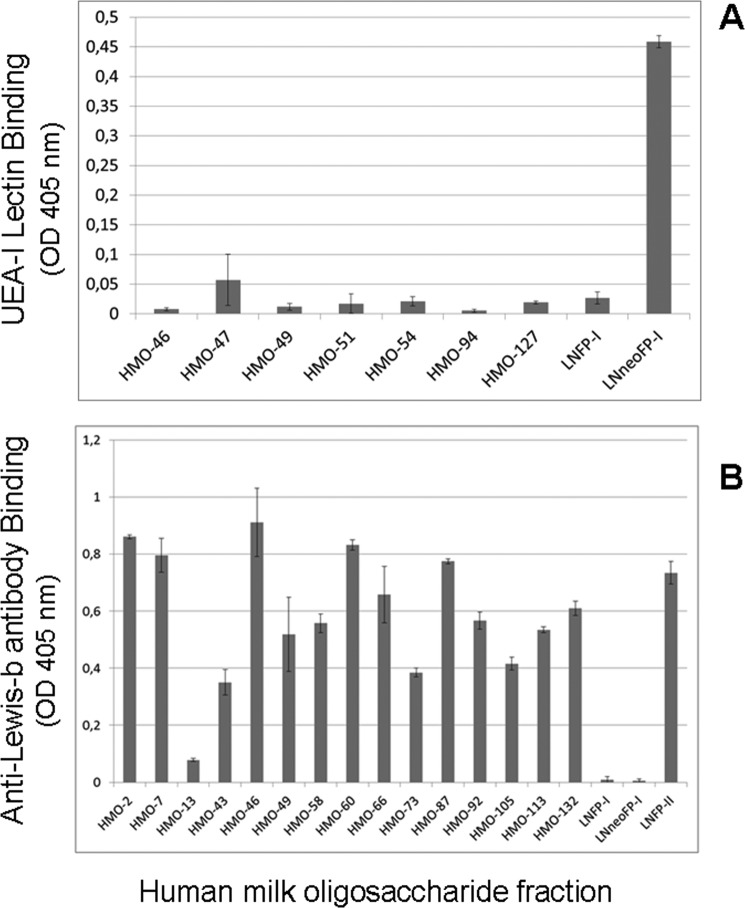
There is agreement with respect to norovirus infection routes in humans regarding binding of the pathogen to gastrointestinal epithelia via recognition of blood group-active mucin-typeO-glycans as the initiating and essential event. Among food additives playing a potential role in applications to protect newborns, human milk oligosaccharides (HMOs) as competitors are of major importance. By focusing on fractions of high-molecular mass HMOs with high fucose contents, we attempted to identify the structural elements required for norovirus GII.4 (Sydney 2012, JX459908) capsid binding in neoglycolipid-based arrays. We provide evidence that HMO fractions with the strongest binding capacities contained hepta- to decasaccharides expressing branches with terminal blood group H1 or Lewis-b antigen. H2 antigen, as recognized by UEA-I lectin, is apparently not expressed in high-mass HMOs. Beyond affinity, sterical and valency effects contribute more to virus-like particle binding, as revealed for oligovalent fucose conjugates of α-cyclodextrin and oligofucoses from fucoidan. Accordingly, high-mass HMOs with oligovalent fucose can exhibit stronger binding capacities compared with monovalent fucose HMOs. The above features were revealed for the most clinically relevant and prevalent GII.4 strain and are distinct from other strains, like GII.10 (Vietnam 026, AF504671), which showed a preference for blood group Lewis-a positive glycans.

Keywords: anti-viral protection; carbohydrate-binding protein; food additive; glycobiology; human blood group antigen; innate immunity; milk oligosaccharide; norovirus; oligosaccharide; virus.


Conflict of interest statement

The authors declare that they have no conflicts of interest with the contents of this article

Figures

Figure 1.
Figure 1.
Binding of norovirus GII.4 (Sydney, 2012, JX459908) VLPs on neoglycolipid arrays derived from high-mass human milk oligosaccharides. A, neoglycolipid fractions 1–42 correspond to HMO fractions listed in Table S1. Two micrograms of neoglycolipid were immobilized per well in a duplicate assay on polystyrene plates. B, neoglycolipids derived from standard HMOs (LNFP-I, lacto-N-fucopentaose-I; LNneoFP-I, lacto-N-neofucopentaose-I; LNFP-II, lacto-N-fucopentaose-II) and selected HMO fractions were tested in triplicate for comparative norovirus GII.4 VLP binding.
Figure 2.
Figure 2.
Comparative profiling of selected HMO fractions as neoglycolipids for blood group H2 and Lewis-b activity. A, binding pattern of U. europaeus lectin isoform I to neoglycolipids derived from selected HMO fractions. Triplicate assay on 2 μg of immobilized neoglycolipids per well. B, binding pattern of monoclonal anti-Lewis-b antibody (2-25LE) to selected HMO fractions. Triplicate assay on 2 μg of immobilized neoglycolipids per well. LNFP-I, lacto-N-fucopentaose-I; LNneoFP-I, lacto-N-neofucopentaose-I; LNFP-II, lacto-N-fucopentaose-II.
Figure 3.
Figure 3.
Inhibition of norovirus GII.4 (Sydney, 2012, JX459908) VLP binding to porcine gastric mucins by HMOs, 2′FL and LNFP-I. A triplicate assay on immobilized porcine stomach mucins (10 μg/ml) was performed. Plates were developed with anti-rabbit Ig-HRP/OPD and read at 490 nm.
Figure 4.
Figure 4.
Binding profiles and structural features of oligosaccharides in HMO fraction 46. A, chromatographic profile of oligosaccharides in HMO fraction 46 (upper panel) and duplicate assay of norovirus GII.4 (Sydney, 2012, JX459908) VLP binding to HPLC subfractions 1–12 (lower panel). B, MALDI-MS survey spectrum of component oligosaccharides in fraction HPLC-7 (upper panel) and MALDI-MS/MS spectrum of methylated glycan with M + Na at m/z 1898 (lower panel). Fragmentation is annotated according to the nomenclature of Domon and Costello (30).
Figure 5.
Figure 5.
Linkage analysis of oligosaccharides in fraction 46 by GC-MS of partially methylated alditol acetates. The ion traces at m/z 118 (a fragment characterizing most of the hexoses) and m/z 159 (fragment characterizing N-acetylhexosamine alditols) are shown.
Figure 6.
Figure 6.
Binding analysis of norovirus GII.4 (Sydney, 2012, JX459908) and GII.10 (Vietnam 026, AF504671) VLPs to human gastric mucins from blood group Lewis-defined individuals. A, binding of norovirus GII.4 VLPs to human gastric mucins from blood group Lewis-defined individuals (triplicate assay); the sample derived from individual Lewis-a positive gastric juice corresponds to sample HGM-Le-a (bar 1) in B. B, binding of norovirus GII.10 VLPs to human gastric mucins from blood group Lewis-defined individuals (triplicate assay); gastric juice from three independent individuals of blood group Lewis-a were tested (HGM-Le-a (1–3)).
Figure 7.
Figure 7.
Inhibition of norovirus GII.4 (Sydney, 2012, JX459908) VLP binding to human gastric mucins by native and processed fucoidan from F. vesiculosus. A, VLP binding assay (triplicate). B, methylation analysis of processed fucoidan. C, positive-ion MALDI mass spectrometry of processed fucoidan (dp, degree of polymerization).
Figure 8.
Figure 8.
Inhibition of norovirus GII.4 (Sydney, 2012, JX459908) VLP binding to human gastric mucins by α-fucosyl–cyclodextrin dendrimers. A triplicate assay of inhibitory potential of FCD1–FCD3 at fixed concentrations was performed: 13.4 mm (FCD1), 11.1 mm (FCD2), and 4.8 mm (FCD3).
Figure 9.
Figure 9.
Structural characterization of α-fucosyl–cyclodextrin dendrimers by MS and linkage analysis by GC-MS. A, MALDI-MS survey spectrum of native α-fucosyl–cyclodextrin dendrimer preparation FCD3. F refers to the mass increment of deoxyhexose (dHex). Up to six dHex residues were added to the scaffold by acid reversion with an average of about three residues. B, GC-MS analysis of partially methylated alditol acetates derived from FCD3. Three major signals were detected at ion traces m/z 175 (terminal Fuc at 8.8 min) and m/z 118 (4-Glc and 4,6-Glc at 11.2 and 12.3 min, respectively). The ratio of 4,6-Glc and 4-Glc in the ion trace at m/z 118 indicates that at least one-third of the available sites on the cyclodextrin scaffold is substituted with fucose. Minor or trace components were registered in the m/z 190 trace at 9.9 min (3-Fuc) and 11.9 min (2,4-Glc), which correspond to less than 2% of the base-peak area (4-Glc) in the TIC chromatogram. a.u., arbitrary unit.

References

    1. van Beek J., Ambert-Balay K., Botteldoorn N., Eden J. S., Fonager J., Hewitt J., Iritani N., Kroneman A., Vennema H., Vinjé J., White P. A., Koopmans M., and NoroNet. (2013) Indications for worldwide increased norovirus activity associated with emergence of a new variant of genotype II.4, late 2012. Euro Surveill. 18, 8–9 - PubMed
    1. Prasad B. V., Hardy M. E., Dokland T., Bella J., Rossmann M. G., and Estes M. K. (1999) X-ray crystallographic structure of the Norwalk virus capsid. Science 286, 287–290 10.1126/science.286.5438.287 - DOI - PubMed
    1. Zheng D. P., Ando T., Fankhauser R. L., Beard R. S., Glass R. I., and Monroe S. S. (2006) Norovirus classification and proposed strain nomenclature. Virology 346, 312–323 10.1016/j.virol.2005.11.015 - DOI - PubMed
    1. Fankhauser R. L., Monroe S. S., Noel J. S., Humphrey C. D., Bresee J. S., Parashar U. D., Ando T., and Glass R. I. (2002) Epidemiologic and molecular trends of “Norwalk-like Viruses” associated with outbreaks of gastroenteritis in the United States. J. Infect. Dis. 186, 1–7 10.1086/341085 - DOI - PubMed
    1. Marionneau S., Ruvoën N., Le Moullac-Vaidye B., Clement M., Cailleau-Thomas A., Ruiz-Palacois G., Huang P., Jiang X., and Le Pendu J. (2002) Norwalk virus binds to histo-blood group antigens present on gastroduodenal epithelial cells of secretor individuals. Gastroenterology 122, 1967–1977 10.1053/gast.2002.33661 - DOI - PMC - PubMed

Publication types