Review
. 2018 Jun 1;10(6):a022608.
doi: 10.1101/cshperspect.a022608.

Microtubules and Microtubule-Associated Proteins

Affiliations
Review

Microtubules and Microtubule-Associated Proteins

Holly V Goodson et al. Cold Spring Harb Perspect Biol. .

Abstract

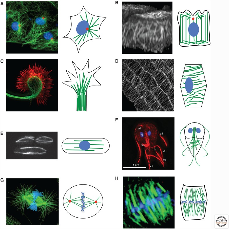
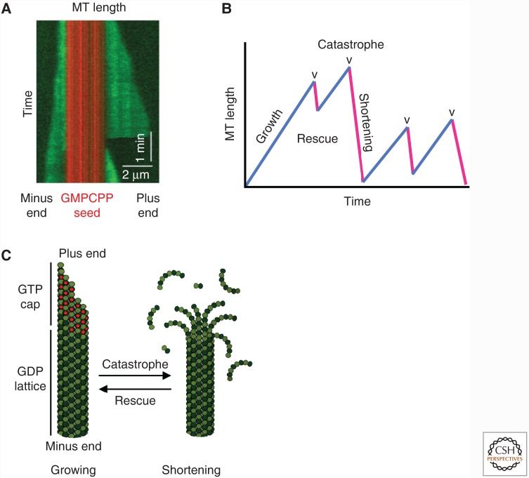
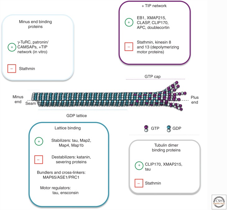
Microtubules act as "railways" for motor-driven intracellular transport, interact with accessory proteins to assemble into larger structures such as the mitotic spindle, and provide an organizational framework to the rest of the cell. Key to these functions is the fact that microtubules are "dynamic." As with actin, the polymer dynamics are driven by nucleotide hydrolysis and influenced by a host of specialized regulatory proteins, including microtubule-associated proteins. However, microtubule turnover involves a surprising behavior-termed dynamic instability-in which individual polymers switch stochastically between growth and depolymerization. Dynamic instability allows microtubules to explore intracellular space and remodel in response to intracellular and extracellular cues. Here, we review how such instability is central to the assembly of many microtubule-based structures and to the robust functioning of the microtubule cytoskeleton.


Figures

Figure 1.
Figure 1.
The microtubule cytoskeleton in various cell types. Each pair of panels contains a fluorescence microscopy image of a specific cell/group of cells (left) with a cartoon depicting the generalized microtubule organization in that cell type (right). The color schemes for the microscope images are described below. In the cartoons, microtubules are shown in green, the DNA in blue, and centrosomes in red. Noncentrosomal microtubule nucleation machinery exists in many cell types (see text) but is not depicted. (A) Radial microtubule array in interphase cells. Microtubules (green), DNA (blue), microtubule-organizing center (MTOC; red). (B) Columnar microtubule array in polarized epithelial cells (green-fluorescent protein [GFP]–tubulin expressed in Madin–Darby canine kidney [MDCK] cells). (C) Microtubules in a neuronal growth cone. Microtubules (green) and actin filaments (red). (D) Cortical microtubule array in plant cells (GFP–tubulin expressed in Arabidopsis cells). (E) Fission yeast interphase microtubules (GFP–tubulin). (F) Microtubule cytoskeleton in Giardia. Microtubules (red), DNA (blue). (G) Animal cell mitotic spindle. Microtubules (green) and DNA (blue). (H) Metaphase plant mitotic spindle. Microtubules (green) and DNA (blue). (A, Reproduced from Gundersen laboratory website [http://www.columbia.edu/~wc2383/pictures.html]; B, reprinted from Reilein et al. 2005; C, reprinted from Kalil et al. 2011; D, reprinted from Ehrhardt and Shaw 2006, with permission of Annual Review of Plant Biology; E, reprinted, with permission, from Chang and Martin 2009, © Cold Spring Harbor Laboratory Press; F, reprinted, with permission, from Dawson 2010, © John Wiley & Sons Inc.; G, left image, reprinted from O’Connell and Khodjakov 2007, with permission from Elsevier, originally from Cell Motility and Cytoskeleton [1999] V.43[3] [cover], with permission from Wiley; H, reproduced from Yu et al. 1999.)
Figure 2.
Figure 2.
Microtubule structure. (A,B) Key aspects of microtubule structure, as indicated. (C) Diagram of the relationship between protofilament number and microtubule structure. (D) Model of the γ-tubulin ring complex (γ-TuRC) associated with the minus end of a microtubule (gray). MTOC, microtubule-organizing center. (A,B, Modified from Kollman et al. 2011, with permission from Macmillan Publishers; C, modified, with permission, from Amos 2004, with permission of The Royal Society of Chemistry, http://dx.doi.org/10.1039/B403634D.)
Figure 3.
Figure 3.
Microtubule (MT) dynamics and assembly. (A) Kymograph (length/time plot derived from a movie) of a microtubule undergoing dynamic instability in vitro, with dynamics at both the minus (left) and plus (right) ends. Green represents Alexa488-labeled tubulin, and red represents tetra-rhodamine-labeled tubulin GMPCPP (guanylyl 5′-α,β-methylenediphosphonate)-stabilized microtubule seeds. (B) Cartoon of a length-history plot (also called a life-history plot) of a microtubule undergoing dynamic instability. The key processes of microtubule dynamics are indicated. (C) Standard model of dynamic instability. As long as the microtubule has a GTP cap, it can grow, but it transitions to rapid depolymerization (catastrophe) on loss of the GTP cap. (A, Adapted, with permission, from Zanic et al. 2013.)
Figure 4.
Figure 4.
Microtubule-binding proteins. Model summarizing some of the major microtubule-binding proteins according to their localization on the microtubule and their activities. The green plus symbol (+) means positive regulation and the red minus symbol (–) means negative regulation. At the plus end (fast growing), the members of the +TIP network (EB1, XMAP215, CLASP, CLIP170, doublecortin, and others not shown) associate with the stabilizing (GTP or GDP-Pi) cap of the growing microtubule and stabilize this dynamic structure to promote growth. Conversely, proteins such as the depolymerizing kinesins and stathmin facilitate microtubule disassembly. At the minus end (slow growing), proteins such as γ-TURC and Patronin/CAMSAP associate with the α-tubulin subunit to cap the end of the filament to prevent depolymerization, which is promoted by stathmin. In the central part of the microtubule, the GDP microtubule lattice can be stabilized by the activities of classical MAPs (tau, Map2, Map4, stop proteins) or destabilized by severing proteins (e.g., katanin). Microtubule-binding proteins that regulate the activity of microtubule motors also bind along the GDP lattice. Microtubules can form large networks through the activities of bundlers/cross-linkers, such as MAP65/ASE1/PRC1. Tubulin dimer binding proteins include stathmin (which promotes depolymerization by sequestering tubulin), as well as CLIP-170, tau, and XMAP-215 (which promote polymerization). Further detail and discussion are available in the main text and references.

References

    1. Ada R, Kilmartin JV. 2000. Spindle pole body duplication: A model for centrosome duplication? Trends Cell Biol 10: 329–335. - PubMed
    1. Akhmanova A, Hoogenraad CC. 2015. Microtubule minus-end-targeting proteins. Curr Biol 25: R162–R171. - PubMed
    1. Akhmanova A, Steinmetz MO. 2008. Tracking the ends: A dynamic protein network controls the fate of microtubule tips. Nat Rev Mol Cell Biol 9: 309–322. - PubMed
    1. Akhmanova A, Steinmetz MO. 2015. Control of microtubule organization and dynamics: Two ends in the limelight. Nat Rev Mol Cell Biol 16: 711–726. - PubMed
    1. Akhmanova A, Stehbens SJ, Yap AS. 2009. Touch, grasp, deliver and control: Functional cross-talk between microtubules and cell adhesions. Traffic 10: 268–274. - PubMed

Publication types

MeSH terms

Substances