. 2018 Jun 1;16(1):148.
doi: 10.1186/s12967-018-1524-5.

Selective pharmacological inhibition of DDR1 prevents experimentally-induced glomerulonephritis in prevention and therapeutic regime

Affiliations

Selective pharmacological inhibition of DDR1 prevents experimentally-induced glomerulonephritis in prevention and therapeutic regime

Solange Moll et al. J Transl Med. .

Abstract

Background: Discoidin domain receptor 1 (DDR1) is a collagen-activated receptor tyrosine kinase extensively implicated in diseases such as cancer, atherosclerosis and fibrosis. Multiple preclinical studies, performed using either a gene deletion or a gene silencing approaches, have shown this receptor being a major driver target of fibrosis and glomerulosclerosis.

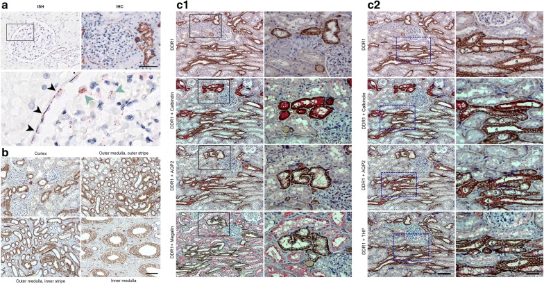
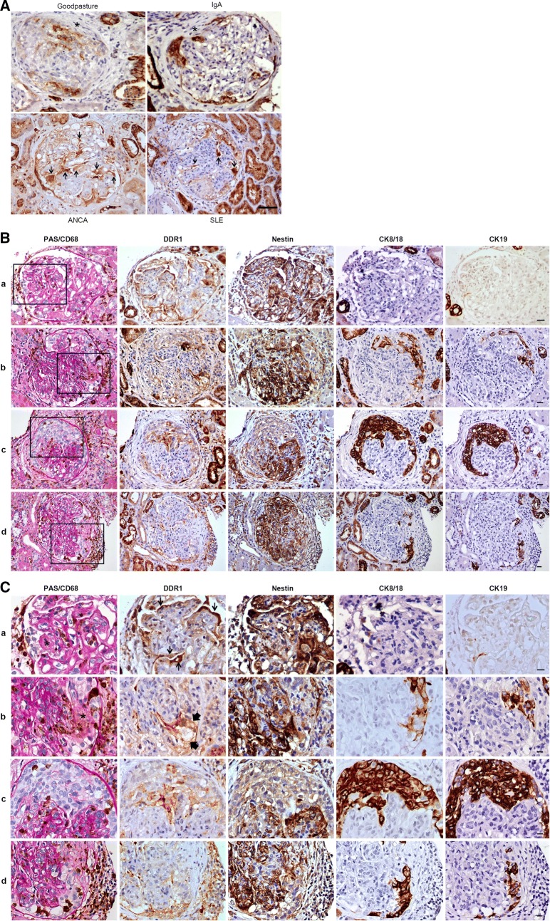
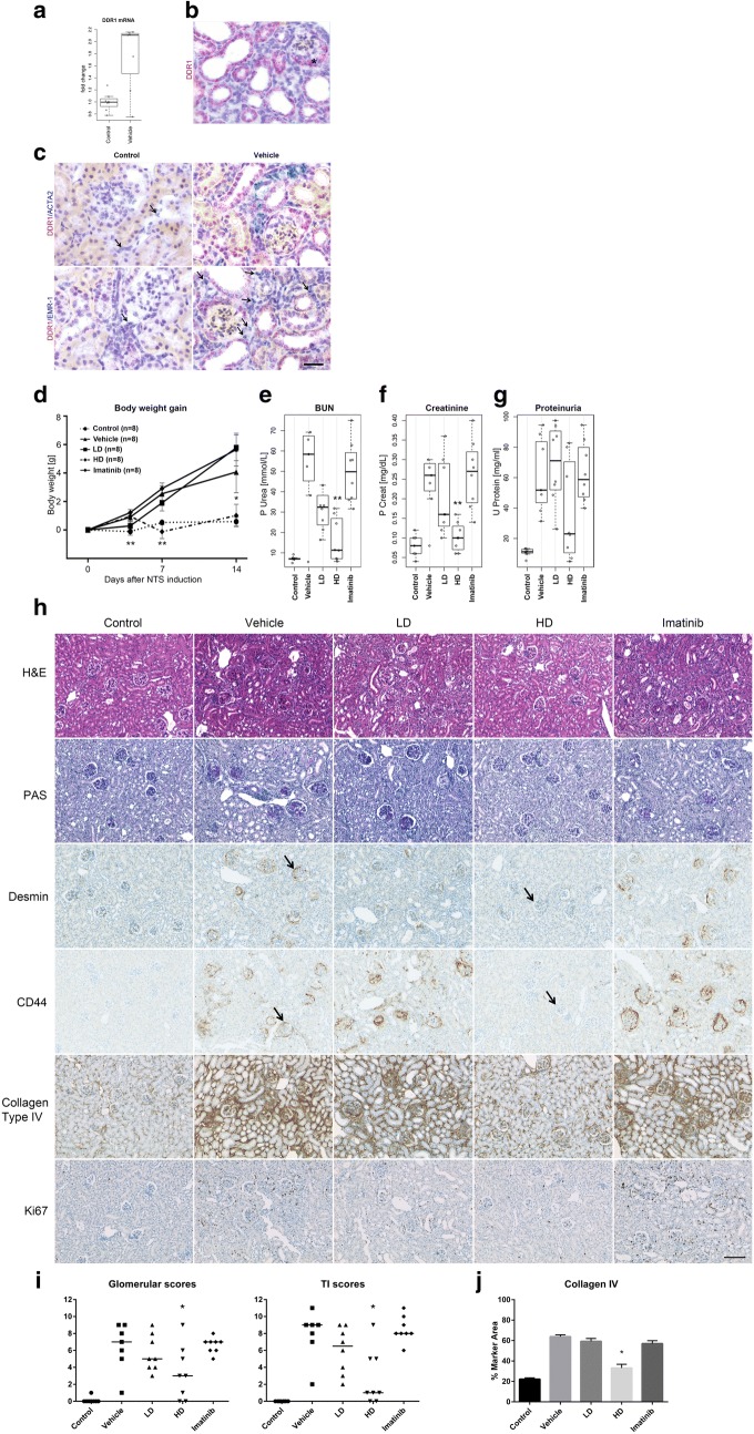
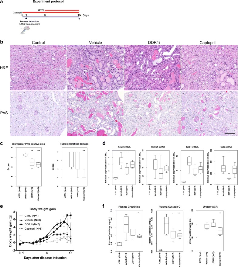
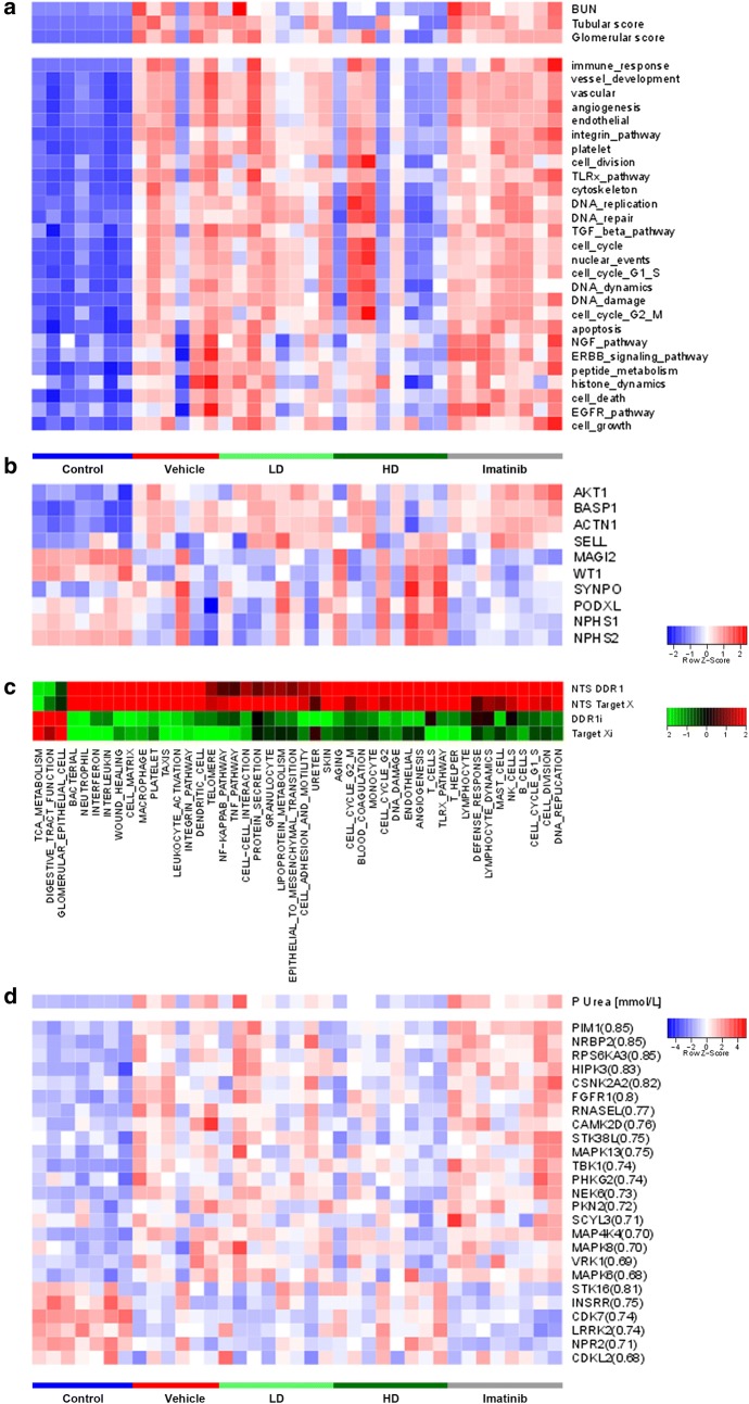
Methods: The present study investigated the role and relevance of DDR1 in human crescentic glomerulonephritis (GN). Detailed DDR1 expression was first characterized in detail in human GN biopsies using a novel selective anti-DDR1 antibody using immunohistochemistry. Subsequently the protective role of DDR1 was investigated using a highly selective, novel, small molecule inhibitor in a nephrotoxic serum (NTS) GN model in a prophylactic regime and in the NEP25 GN mouse model using a therapeutic intervention regime.

Results: DDR1 expression was shown to be mainly limited to renal epithelium. In humans, DDR1 is highly induced in injured podocytes, in bridging cells expressing both parietal epithelial cell (PEC) and podocyte markers and in a subset of PECs forming the cellular crescents in human GN. Pharmacological inhibition of DDR1 in NTS improved both renal function and histological parameters. These results, obtained using a prophylactic regime, were confirmed in the NEP25 GN mouse model using a therapeutic intervention regime. Gene expression analysis of NTS showed that pharmacological blockade of DDR1 specifically reverted fibrotic and inflammatory gene networks and modulated expression of the glomerular cell gene signature, further validating DDR1 as a major mediator of cell fate in podocytes and PECs.

Conclusions: Together, these results suggest that DDR1 inhibition might be an attractive and promising pharmacological intervention for the treatment of GN, predominantly by targeting the renal epithelium.

Keywords: CKD; DDR1 inhibition; Fibrosis; Glomerulosclerosis.


Figures

Fig. 1
Fig. 1
Localisation of DDR1 in human control kidney. a DDR1 in situ hybridization (ISH) and immunohistochemistry (IHC) analyses in normal human kidney. (Scale bar = 100 μm). ISH higher magnification rectangle: DDR1 positive podocyte (black arrowheads), DDR1 positive podocytes (green arrowheads). b DDR1 immunostaining in the nomal human kidney with representative images of the cortex, outer medulla (outer and inner stripes) and inner medulla. Note the DDR1 membranous staining of the tubules. (Magnification ×125, scale bar 50 μm). c Serial sections of normal human kidney were double immunostained with DDR1 and specific tubular antibodies against Megalin, Calbindin, Tamm–Horsfall Protein (THP or Uromodulin) or Aquaporin 2 (AQP2). c1 Representative micrographs of the cortex show DDR1 protein localization in the distal convoluted tubules (Calbindin+, Aquaporin 2−, Megalin−). Proximal tubules (Megalin+) are DDR1 negatively stained. Boxed areas are enlarged in the right side of the figure. c2 Representative micrographs of the medullar rays of the cortex show DDR1 protein localization in the connecting tubules (1: Calbindin+, Aquaporin 2+, THP−), distal convoluted tubules (2: Calbindin+, Aquaporin 2−, THP−), thick ascending limbs of the Henle’s loop/pars recta of the distal tubules (3: THP+, Calbindin−, Aquaporin 2−) and collecting ducts (4: Aquaporin 2+, Calbindin−, THP−). Boxed areas are enlarged in the right side of the figure. Magnification ×100, scale bar = 100 μm; Boxed areas Magnification ×250, scale bar = 50 μm
Fig. 2
Fig. 2
DDR1 is highly induced in crescentic glomerulonephritis. A DDR1 expression in 4 different types of human crescentic glomerulonephritis. DDR1 immunostaining in human kidney biopsies from patients with 4 different types of crescentic glomerulonephritis (GN): Goodpasture’s syndrome (patient #1), ANCA-associated GN (patient #12), IgA GN (patient #19) and lupus GN class IV-G (A/C) (patient #29). DDR1 is expressed in the cellular crescents of all 4 types of crescentic GN with variability in the staining intensity and expression limited within crescents to a subset of cells. Injured podocytes and bridging podocytes (arrows) strongly express DDR1. Fibrous crescent (*) are DDR1 negative. Magnification ×200 for SLE, ×250 for ANCA, ×320 for Goopasture and IgA, Scale bar = 20 μm. BC Detailed analysis of DDR1 expresion during crescent formation. Serial sections of human kidney biopsies from patients with lupus GN class IV-G (A/C) (patients #25 and #29) were immunostained with specific antibodies against CD68/PAS (Periodic Acid Schiff), DDR1, nestin, cytokeratin 8/18 or cytokeratin 19 proteins. B Representative micrographs show expression and localization of each protein in the glomeruli. Boxed areas represent crescentic lesion and are enlarged in C. Lines a–d illustrate 4 different morphological stages of the crescent formation; a: early stage with fibrin within the glomerular capillary lumen and presence of 2–3 layers of cells in Bowman’s space; b: early stage with gap in the glomerular capillary wall, plasma proteins in Bowman’s space and cellular crescent formation; c: active hypercellular circumferential crescent compressing the glomerular tuft; d: advanced stage with fibrocellular crescent, capsular rupture and periglomerular inflammation. Line a = patient #25; lines b–d = patient #29. Fine arrows = injured podocytes; large arrows = bridging cells; * = fibrin and plasma proteins within the glomerular capillary lumen or in Bowman’s space. Magnification A line a: ×320, b and c: ×250, d: ×200; B line a: ×640, b and c: ×500 d: ×400; Scale bar = 20 μm
Fig. 3
Fig. 3
Pharmacological inhibition of DDR1 activation protects animals against NTS-induced crescentic glomerulonephritis. a Quantitative RT-PCR for Ddr1 mRNA on whole kidney lysate of control mice (Control) and mice injected with nephrotoxic serum and treated with vehicle (vehicle). b Representative Ddr1 in situ hybridization (ISH) performed on tissue harvested from mice 14 days after NTS injection. * = crescent c representative DDR1 ISH double labelling with alpha smooth muscle actin (Acta2) or EGF-like module-containing mucin-like hormone receptor-like 1 (Emr-1) in control mice (Control) and mice injected with nephrotoxic serum and treated with vehicle (Vehicle). Arrows = cells labeled with Acta2 or Emr-1. dg Body weight evolution (day 1, 4, 7 and 14) and renal function parameters (eg blood urea nitrogen (BUN), serum creatinine and proteinuria) measured at sacrifice (day 14). h Representative histopathology with Hematoxylin and Eosin (H&E) and Periodic Acid Schiff staining (PAS) and immunohistochemistry for desmin, CD44, Collagen type IV and Ki67. i Glomerular or tubulo-interstitial (TI) summary scores from semiquantitative histopathologic evaluation on H&E and PAS stained kidney sections respectively. j Morphometry analysis of collagen IV IHC. Statistically significant p value: p < 0.05 = *; p < 0.005 = **. Magnification ×200, scale bar 100 μm
Fig. 4
Fig. 4
Treatment with DDR1 inhibitor in the NEP25 mouse model of glomerulosclerosis. a Schema of the experiment b Representative histopathology with Hematoxylin and Eosin (H&E) and Periodic Acid Schiff staining (PAS) in control, vehicle-, DDR1i- and Captopril treated groups at day 15. Magnification ×200, scale bar 100 μm. c Semi-quantificative analysis of glomerulosclerosis (glomerular PAS positive area) and tubulointerstitial lesions (tubulointerstitial damage) in control, vehicle-, DDR1i- and Captopril treated groups at day 15. ***p < 0.001, ****p < 0.0001; t-test and Mann–Whitney U test were used for the score of glomerular PAS positive area and tubulointerstitial damage respectively. d Quantitative RT-PCR for the fibrosis markers alpha smooth muscle actin (Acta2 mRNA), collagen type 1 (Col1a mRNA) and TGF-β1 (TGF-β1 mRNA), and for the inflammation marker Ccl2 in NEP25 mice treated with DDR1 inhibitor (DDR1i), Captopril or vehicle and in control mice. *p < 0.05, **p < 0.01, ***p < 0.001; t-test. e Body weight evolution. f Renal function parameters (plasma creatinine and plasma Cystatin C measured at sacrifice) and urinary ACR (24-h urine collection from day 14 to 15 divided by creatinine concentration) in NEP25 mice treated with DDR1 inhibitor (DDR1i), Captopril or vehicle and in control mice (CTRL)
Fig. 5
Fig. 5
Gene expression profile in NTS-treated mice. a Unsupervised gene signature analysis and GSEA using Gene Ontology processes as input genesets. b Glomerular cell signature with individual component genes. c Comparison of different target modulation in NTS. Heatmap showing differential modulation (by DDR1i or Target X) of the gene networks modulated by NTS induction compared to control. d Heatmap showing kinome expression modulation in the different treatment conditions

Similar articles

Cited by

References

    1. Vogel W, et al. The discoidin domain receptor tyrosine kinases are activated by collagen. Mol Cell. 1997;1(1):13–23. doi: 10.1016/S1097-2765(00)80003-9. - DOI - PubMed
    1. Valiathan RR, et al. Discoidin domain receptor tyrosine kinases: new players in cancer progression. Cancer Metastasis Rev. 2012;31(1–2):295–321. doi: 10.1007/s10555-012-9346-z. - DOI - PMC - PubMed
    1. Leitinger B. Discoidin domain receptor functions in physiological and pathological conditions. Int Rev Cell Mol Biol. 2014;310:39–87. doi: 10.1016/B978-0-12-800180-6.00002-5. - DOI - PMC - PubMed
    1. Borza CM, Pozzi A. Discoidin domain receptors in disease. Matrix Biol. 2014;34:185–192. doi: 10.1016/j.matbio.2013.12.002. - DOI - PMC - PubMed
    1. Yeh YC, Lin HH, Tang MJ. A tale of two collagen receptors, integrin beta1 and discoidin domain receptor 1, in epithelial cell differentiation. Am J Physiol Cell Physiol. 2012;303(12):C1207–C1217. doi: 10.1152/ajpcell.00253.2012. - DOI - PubMed

MeSH terms

Substances