Review
. 2018 Oct:51:116-124.
doi: 10.1016/j.yfrne.2018.05.004. Epub 2018 May 31.

TRPCing around the hypothalamus

Affiliations
Review

TRPCing around the hypothalamus

Martin J Kelly et al. Front Neuroendocrinol. 2018 Oct.

Abstract

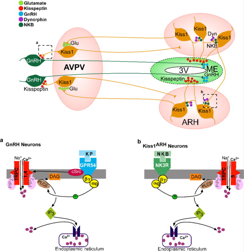
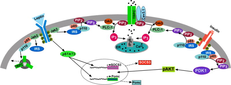
All of the canonical transient receptor potential channels (TRPC) with the exception of TRPC 2 are expressed in hypothalamic neurons and are involved in multiple homeostatic functions. Although the metabotropic glutamate receptors have been shown to be coupled to TRPC channel activation in cortical and sub-cortical brain regions, in the hypothalamus multiple amine and peptidergic G protein-coupled receptors (GPCRs) and growth factor/cytokine receptors are linked to activation of TRPC channels that are vital for reproduction, temperature regulation, arousal and energy homeostasis. In addition to the neurotransmitters, circulating hormones like insulin and leptin through their cognate receptors activate TRPC channels in POMC neurons. Many of the post-synaptic effects of the neurotransmitters and hormones are regulated in different physiological states by expression of TRPC channels in the post-synaptic neurons. Therefore, TRPC channels are key targets not only for neurotransmitters but circulating hormones in their vital role to control multiple hypothalamic functions, which is the focus of this review.

Keywords: 17β-estradiol; GnRH; Insulin; Kisspeptin; Leptin; Neurokinin B; Orexin; POMC; TRPC channels.


Conflict of interest statement

Disclosure Statement

The authors have nothing to disclose.

Figures

Figure 1
Figure 1. A model by which activation of Kiss1 neurons governs GnRH neuronal excitability through TRPC channels
Activation of Kiss1 neurons in the ARH releases neurokinin B (NKB) that depolarizes and recruits other Kiss1ARH neurons. Dynorphin is co-released and acts presynaptically to modulate (inhibit) the release of NKB. Together the two peptides govern the synchronous activity of Kiss1ARH neurons and promote kisspeptin release that stimulates GnRH release in the median eminence (ME). Kiss1ARH neurons also communicate with the Kiss1AVPV/PeN neurons via the fast neurotransmitter glutamate, which stimulates burst-firing of Kiss1AVPV/PeN neurons. Activation of these rostral Kiss1 neurons releases kisspeptin to robustly excite GnRH neurons via activation of the GPR54 (Kiss1R) signaling cascade, thereby stimulating the release of GnRH at the time of the preovulatory surge. Kisspeptin, GPR54, NKB, TacR3 and GnRH are all required for normal fertility. [Modified from Fig. 12, Qiu et al., 2016. Elife doi: 10.7554/eLife.16246.] (a) Kisspeptin excites GnRH neurons through TRPC 4 channels. Kisspeptin binds to the Gq-coupled GPR54 receptor to activate phospholipase Cβ (PLCβ), which catabolizes PIP2, potentiates TRPC channel activity and inhibits the inwardly rectifying K+ channel activity. cSRC kinase, which is activated by kisspeptin/GRP54 signaling, potentiates the activity of TRPC 4 channels; and (b) Neurokinin B excites Kiss1ARH neurons through TRPC 5 channel activation.
Figure 2
Figure 2. TacR3 agonist excites Kiss1ARH neurons through TRPC5 channels
(A) TacR3 agonist senktide (500 nM) depolarizes Kiss1ARH neurons in female mice and induces firing (upper trace). In voltage clamp senktide induces an inward current (middle trace). Vhold = −60 mV. The inward current is blocked by TRPC channel blocker 2-aminoethyl diphenylborinate (2-APB; 100 μM) (lower trace). (B) The I-V relationship for the senktide (Senk)-induced inward current reverses at −10 mV and exhibits double rectification. (C) Summary of the effects of TRPC channel blocker 2-APB on the senktide-induced inward currents. The TRPC channel blocker is applied 15 min before the application of senktide (500 nM). Vhold = − 60 mV. ***p < 0.005, different from the senktide control group. Data points represent the mean ± SEM. Unpaired two-tailed t test, t(21) = 4.234, p = 0.0004. Cell numbers tested are indicated. (D) Representative gel images illustrating the mRNA expression of Trpc5 channel subunit in Kiss1ARH neurons harvested from female mice. The expected size of PCR products for Kiss1 and Trpc5 are indicated. MM is the molecular marker; −RT indicates a harvested Kiss1 neuron without RT; + indicates positive tissue control (with RT); – indicates negative tissue control (without RT) using cDNA from mouse medial basal hypothalamic tissue. RT, reverse transcriptase.
Figure 3
Figure 3. A cellular model of insulin (and leptin) signaling via TRPC 5 channel activation in POMC neurons
Insulin signals via IRS-PI3K to activate TRPC 5 channels in POMC neurons, which generates a robust inward cationic current to depolarize POMC neurons and increase their excitability. Similarly, leptin binding to its receptor (LRb) triggers the recruitment of the tyrosine kinase Janus kinase (JAK) 2, leading to the activation of the JAK/signal transducer and activator of transcription (STAT) signaling pathway and simultaneously activation of PI3K, which also activates TRPC 5 channels. PI3K (p85/p110) will also accelerate the rapid insertion of TPRC 5 channels into the plasma membrane. PDK1, 3-phosphoinositide–dependent protein kinase-1. [Modified from Fig. 8, Qiu et al., 2018a. Endocrinology 159, 647-664.]

References

    1. Adamantidis AR, Zhang F, Aravanis AM, Deisseroth K, de Lecea L. Neural substrates of awakening probed with optogenetic control of hypocretin neurons. Nature. 2007;450:420–424. - PMC - PubMed
    1. Al-Qassab H, Smith MA, Irvine EE, Guillermet-Guibert J, Claret M, Choudhury AI, Selman C, Piipari K, Clements M, Lingard S, Chandarana K, Bell JD, Barsh GS, Smith AJH, Batterham RL, Ashford MLJ, Vanhaesebroeck B, Withers DJ. Dominant role of the p110β isoform of PI3K over p110α in energy homeostasis regulation by POMC and AgRP neurons. Cell Metab. 2009;10:343–354. - PMC - PubMed
    1. Ambudkar IS, Ong HL. Organization and function of TRPC channelsomes. Pflügers Arch. 2007;455:187–200. - PubMed
    1. Apergis-Schoute J, Iordanidou P, Faure C, Jego S, Schone C, Aitta-Aho T, Adamantidis A, Burdakov D. Optogenetic evidence for inhibitory signaling from orexin to MCH neurons via local microcircuits. J Neurosci. 2015;35:5435–5441. - PMC - PubMed
    1. Aponte Y, Atasoy D, Sternson SM. AGRP neurons are sufficient to orchestrate feeding behavior rapidly and without training. Nat Neurosci. 2011;14:351–355. - PMC - PubMed

Publication types

MeSH terms