. 2018 Sep:49:59-67.
doi: 10.1016/j.cellsig.2018.05.011. Epub 2018 Jun 1.

Phospholipase C delta 4 (PLCδ4) is a nuclear protein involved in cell proliferation and senescence in mesenchymal stromal stem cells

Affiliations

Phospholipase C delta 4 (PLCδ4) is a nuclear protein involved in cell proliferation and senescence in mesenchymal stromal stem cells

Marianna Kunrath-Lima et al. Cell Signal. 2018 Sep.

Abstract

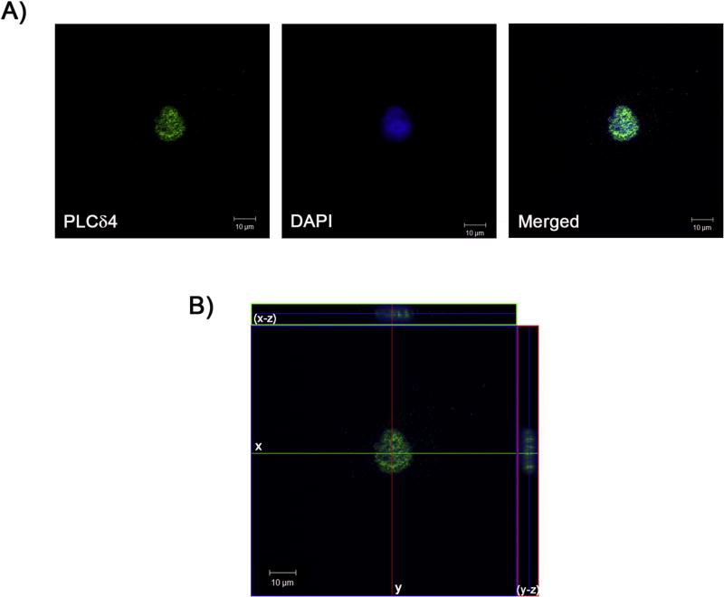
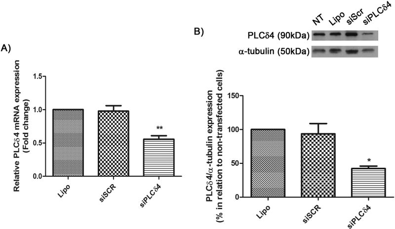
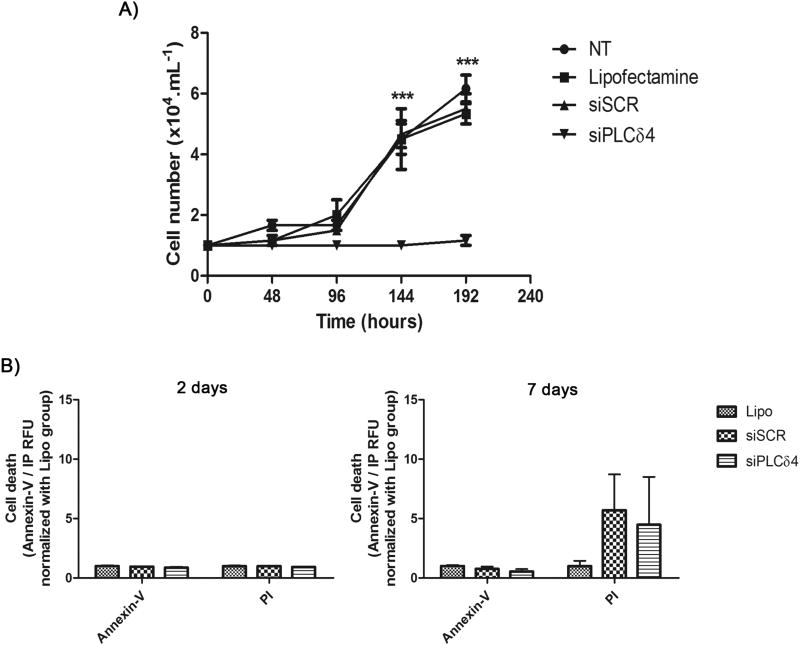
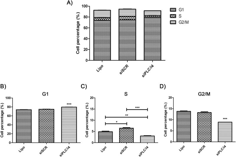
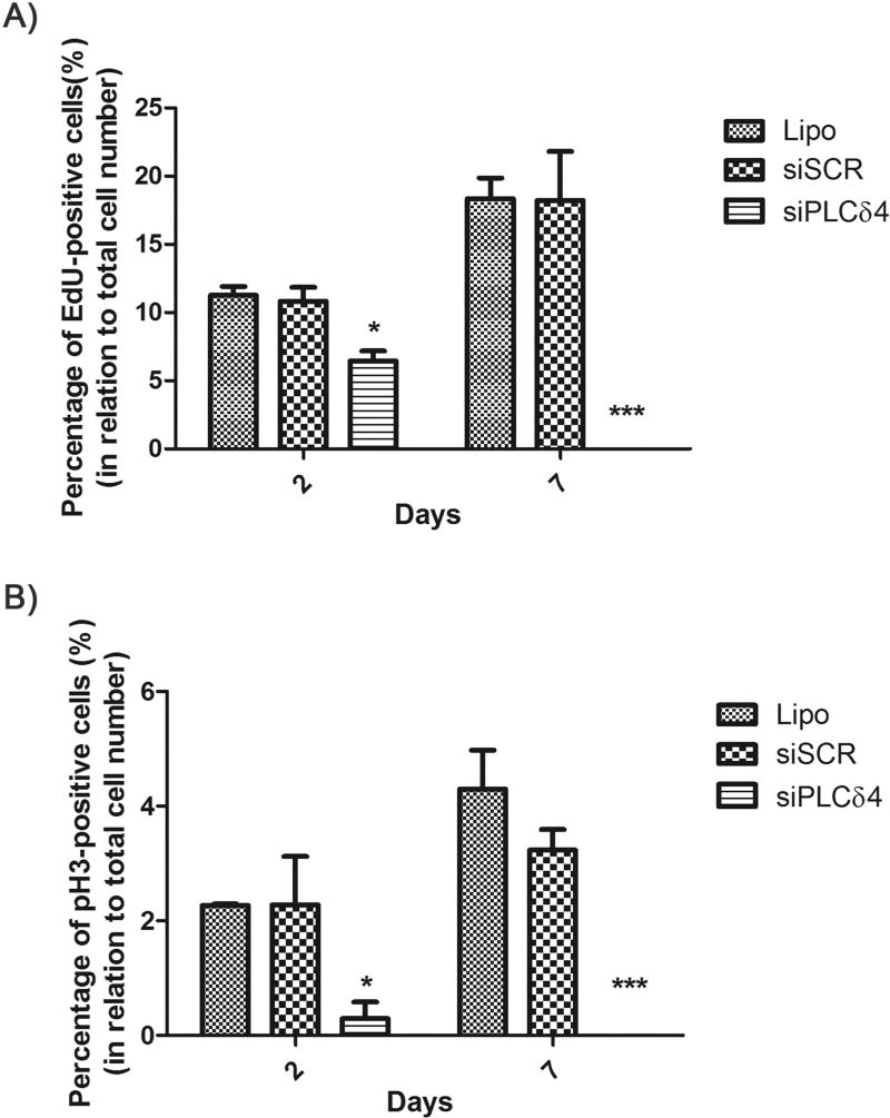
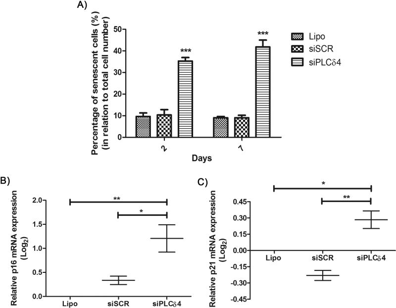
Ca2+ is an important second messenger, and it is involved in many cellular processes such as cell death and proliferation. The rise in intracellular Ca2+ levels can be due to the generation of inositol 1,4,5-trisphosphate (InsP3), which is a product of phosphatidylinositol 4,5-bisphosphate (PIP2) hydrolysis by phospholipases C (PLCs), that leads to Ca2+ release from endoplasmic reticulum by InsP3 receptors (InsP3R). Ca2+ signaling patterns can vary in different regions of the cell and increases in nuclear Ca2+ levels have specific biological effects that differ from those of Ca2+ increase in the cytoplasm. There are PLCs in the cytoplasm and nucleus, but little is known about the functions of nuclear PLCs. This work aimed to characterize phenotypically the human PLCδ4 (hPLCδ4) in mesenchymal stem cells. This nuclear isoform of PLC is present in different cell types and has a possible role in proliferative processes. In this work, hPLCδ4 was found to be mainly nuclear in human adipose-derived mesenchymal stem cells (hASC). PLCδ4 knockdown demonstrated that it is essential for hASC proliferation, without inducing cell death. An increase of cells in G1, and a reduction of cells on interphase and G2/M in knockdown cells were seen. Furthermore, PLCδ4 knockdown increased the percentage of senescent cells, p16INK4A+ and p21Cip1 mRNAs expression, which could explain the impaired cell proliferation. The results show that hPLCδ4 is in involved in cellular proliferation and senescence in hASC.

Keywords: Calcium signaling; Cellular proliferation; Cellular senescence; Human PLCδ4; Nuclear calcium signaling.


Conflict of interest statement

Conflict of interest

The authors declare that they have no competing interests.

Figures

Figure 1
Figure 1. PLCδ4 was primarily nuclear localized in hASC
hASC were incubated with primary antibody anti-PLCδ4 and a secondary antibody conjugated with Alexa 488 (green). The nucleus was stained with DAPI (blue). A) PLCδ4 colocalizes with nuclear staining in hASC, indicated by the merged image. B) Tridimensional reconstruction. Central panel: medial plane inside the nucleus. Upper panel: 3D reconstruction of the x-z plane. Right panel: 3D reconstruction of the y-z plane. Scale bar: 10 µm. Representative image of what was observed in at least samples of three patients.
Figure 2
Figure 2. siPLCδ4 sequence reduces the expression of PLCδ4 in hASC
A) Relative PLCδ4 mRNA expression measured by Real-Time qPCR. Cells were transfected with 25 nM of scrambled siRNA (siSCR) or siRNA for PLCδ4 (siPLCδ4). 48 hours post transfection, total RNA was extracted from cells and was used for cDNA synthesis. PLCδ4 expression was normalized with GAPDH and RPL13A, and samples treated only with lipofectamine (non-transfected cells) were used as calibrators. There was a reduction in PLCδ4 expression in siPLCδ4 cells. B) Western Blot of total protein extracts 48 hours posttransfection. α-tubulin was used as a loading protein control, and protein expression levels were normalized with lipofectamine samples values (in percentage). The reduction of PLCδ4 protein expression can be observed for siPLCδ4 treated cells when compared to controls. Lipo = cells treated with lipofectamine (transfection reagent); siSCR = cells transfected with scrambled siRNA; siPLCδ4 = cells transfected with siRNA for PLCδ4. One-way ANOVA with Bonferroni’s multiple comparison tests (* p<0.05; ** p<0.01, n=3).
Figure 3
Figure 3. PLCδ4 knockdown abolishes cell proliferation in hASC, without a decrease in cell viability
A) Growth curve of transfected cells. The curve was started with 1 × 10⁴ cells. Cells were counted every two days, in a Neubauer chamber, using Trypan Blue for viability exclusion. From 0 to 192 hours the number of cells in siPLCδ4 were stable. The time points of 144 hours and 192 hours in siPLCδ4 are different from the other groups. B) Cell death assay, using Annexin-V/PI. 2 or 7 days post-transfection cells were stained Annexin-V/PI and their fluorescence were measured. There was no difference between the groups. NT = non-treated cells; Lipo = cells treated with lipofectamine (transfection reagent); siSCR = cells transfected with scrambled siRNA; siPLCδ4 = cells transfected with siRNA for PLCδ4. RFU = relative fluorescence unit. Two-way ANOVA with Bonferroni’s multiple comparison tests (*** p<0.001, n=3 for growth curve and n=2 for Annexin-V/PI).
Figure 4
Figure 4. Reduction of PLCδ4 expression in hASC leads to cell cycle arrest
48 hours after transfection, cells were resuspended with trypsin, incubated with the nuclear probe To-Pro® 3 and analyzed by flow cytometry. A) Graphic showing cell cycle phases distribution among the different cell groups. B) Percentage of cells, in relation to total cell number, on G1 cell cycle phase. C) S phase. D) G2/M phases. Lipo = cells treated with lipofectamine (transfection reagent); siSCR = cells transfected with scrambled siRNA; siPLCδ4 = cells transfected with siRNA for PLCδ4. One-way ANOVA with Bonferroni’s multiple comparison tests (* p<0.05; ** p<0.01; *** p<0.001, n≥3).
Figure 5
Figure 5. PLCδ4 knockdown reduces the number of EdU-positive and pH3-positive cells
2 or 7 days after transfection, hASC were incubated with Hoescht (nuclear staining) and EdU (a probe for dividing cells) or pH3 (a marker for mitosis). The figures show the percentage of cells marked simultaneously with Hoechst and EdU or pH3, in relation to total cell number (stained only with Hoescht). A) Percentage of EdU-positive cells. B) Percentage of pH3-positive cells. For both probes and for the two-time points analyzed, there was a reduction of positive-labeled cells for the siPLCδ4 group, when compared to control groups. One-way ANOVA with Bonferroni’s multiple comparison tests (*p<0.05; *** p<0.001, n=3).
Figure 6
Figure 6. PLCδ4 silencing increases the percentage of senescent cells
A) 2 or 7 days after transfection, hASC were assayed for SA-β-Gal activity. The figure shows the percentage of cells stained for SA-β-Gal activity, in relation to total cell number. For the two-time points analyzed, there was a reduction of stained cells for the siPLCδ4 group, when compared to control groups as determined by one-way ANOVA with Bonferroni’s multiple comparison tests (***p<0.001, n=3). B and C) Relative p16INK4A+ and p21Cip1 mRNA expression measured by Real-Time qPCR. 48 hours post transfection, total RNA was extracted from cells and was used for cDNA synthesis. p16 and p21 expression was normalized with RPL13A, and Lipo group was used as calibrator. There was an increase in p16INK4A+ and p21Cip1 expression in siPLCδ4 cells. One-way ANOVA with Bonferroni’s multiple comparison tests (* p<0.05; ** p<0.01, n=2).

References

    1. Berridge MJ, Lipp P, Bootman MD. The versatility and universality of calcium signalling. Nature reviews. Molecular cell biology. 2000;1(1):11–21. - PubMed
    1. Mendes CC, Gomes DA, Thompson M, Souto NC, Goes TS, Goes AM, Rodrigues MA, Gomez MV, Nathanson MH, Leite MF. The type III inositol 1,4,5-trisphosphate receptor preferentially transmits apoptotic Ca2+ signals into mitochondria. The Journal of biological chemistry. 2005;280(49):40892–900. - PubMed
    1. Minagawa N, Nagata J, Shibao K, Masyuk AI, Gomes DA, Rodrigues MA, Lesage G, Akiba Y, Kaunitz JD, Ehrlich BE, Larusso NF, Nathanson MH. Cyclic AMP regulates bicarbonate secretion in cholangiocytes through release of ATP into bile. Gastroenterology. 2007;133(5):1592–602. - PMC - PubMed
    1. Rodrigues MA, Gomes DA, Leite MF, Grant W, Zhang L, Lam W, Cheng YC, Bennett AM, Nathanson MH. Nucleoplasmic calcium is required for cell proliferation. The Journal of biological chemistry. 2007;282(23):17061–8. - PMC - PubMed
    1. Mikoshiba K. The InsP3 receptor and intracellular Ca2+ signaling. Current opinion in neurobiology. 1997;7(3):339–45. - PubMed

MeSH terms

Substances

LinkOut - more resources