

Neural representation of vowel formants in tonotopic auditory cortex
- PMID: 29860083
- PMCID: PMC6231402
- DOI: 10.1016/j.neuroimage.2018.05.072
Neural representation of vowel formants in tonotopic auditory cortex
Abstract
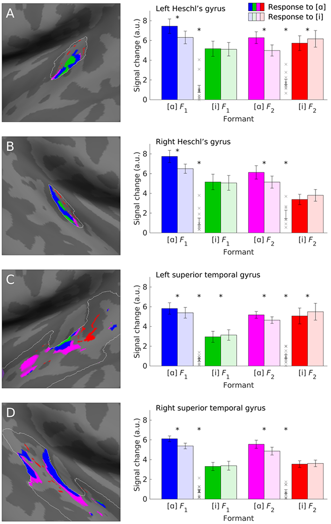
Speech sounds are encoded by distributed patterns of activity in bilateral superior temporal cortex. However, it is unclear whether speech sounds are topographically represented in cortex, or which acoustic or phonetic dimensions might be spatially mapped. Here, using functional MRI, we investigated the potential spatial representation of vowels, which are largely distinguished from one another by the frequencies of their first and second formants, i.e. peaks in their frequency spectra. This allowed us to generate clear hypotheses about the representation of specific vowels in tonotopic regions of auditory cortex. We scanned participants as they listened to multiple natural tokens of the vowels [ɑ] and [i], which we selected because their first and second formants overlap minimally. Formant-based regions of interest were defined for each vowel based on spectral analysis of the vowel stimuli and independently acquired tonotopic maps for each participant. We found that perception of [ɑ] and [i] yielded differential activation of tonotopic regions corresponding to formants of [ɑ] and [i], such that each vowel was associated with increased signal in tonotopic regions corresponding to its own formants. This pattern was observed in Heschl's gyrus and the superior temporal gyrus, in both hemispheres, and for both the first and second formants. Using linear discriminant analysis of mean signal change in formant-based regions of interest, the identity of untrained vowels was predicted with ∼73% accuracy. Our findings show that cortical encoding of vowels is scaffolded on tonotopy, a fundamental organizing principle of auditory cortex that is not language-specific.
Keywords: Auditory cortex; Formants; Tonotopy; Vowels.
Copyright © 2018 Elsevier Inc. All rights reserved.
Figures


References
-
- American Speech-Language-Hearing Association, 1997. Guidelines for audiologic screening. doi:10.1044/policy.GL1997-00199. - DOI
-
- Bates D, Mächler M, Bolker B, Walker S, 2015. Fitting linear mixed-effects models using lme4. J. Stat. Softw. 67, 1–48.
Publication types
MeSH terms
Grants and funding
LinkOut - more resources
Full Text Sources
Other Literature Sources
