. 2018 Jul:17:386-399.
doi: 10.1016/j.redox.2018.04.021. Epub 2018 May 1.

GSNOR modulates hyperhomocysteinemia-induced T cell activation and atherosclerosis by switching Akt S-nitrosylation to phosphorylation

Affiliations

GSNOR modulates hyperhomocysteinemia-induced T cell activation and atherosclerosis by switching Akt S-nitrosylation to phosphorylation

Jing Li et al. Redox Biol. 2018 Jul.

Abstract

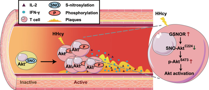
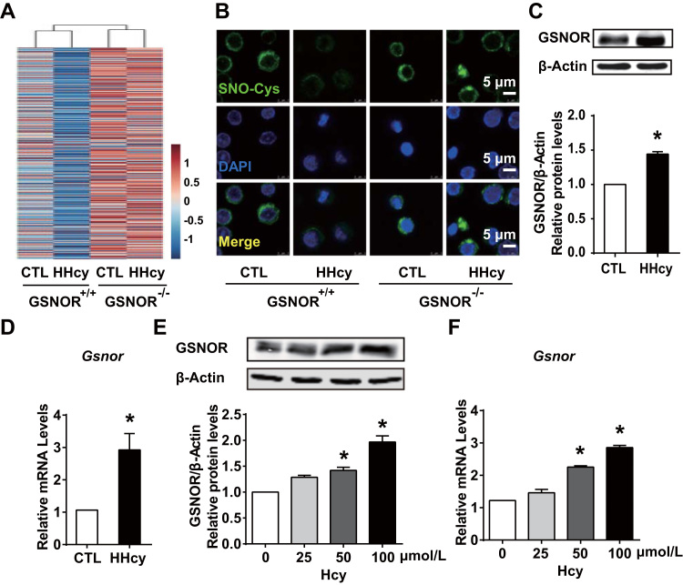
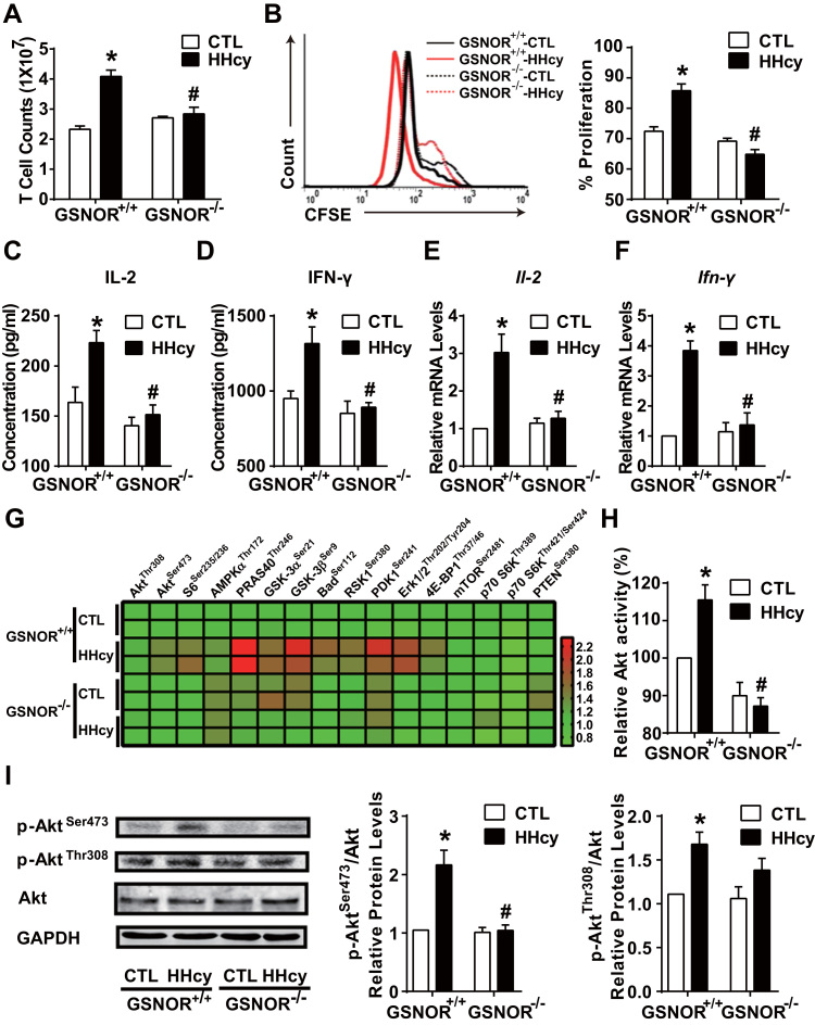
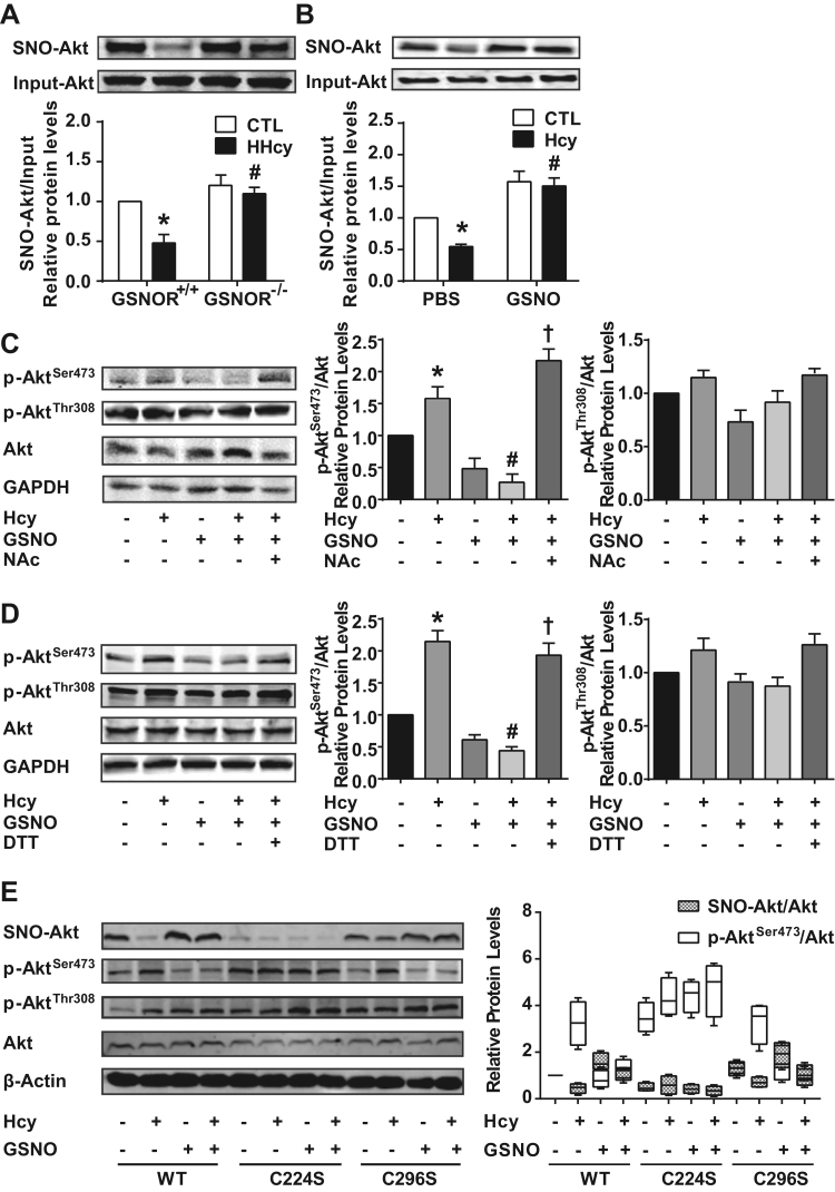
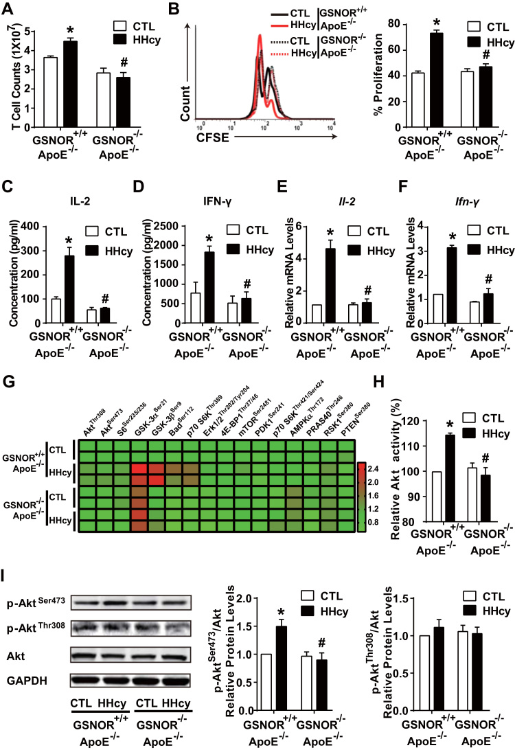
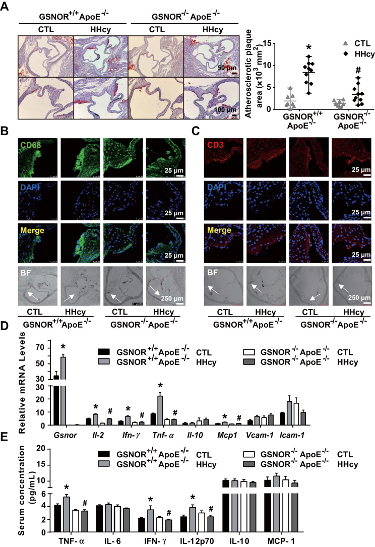
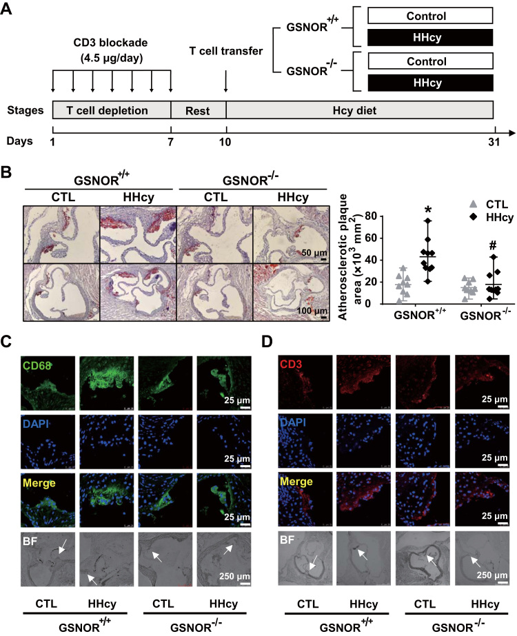
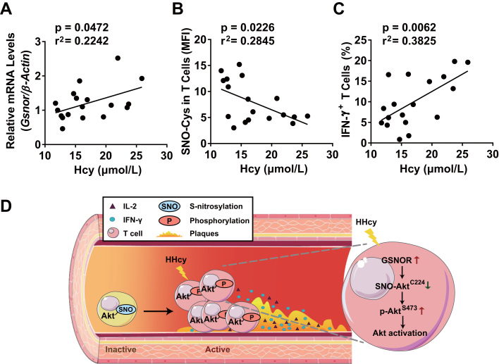
The adaptive immune system plays a critical role in hyperhomocysteinemia (HHcy)-accelerated atherosclerosis. Recent studies suggest that HHcy aggravates atherosclerosis with elevated oxidative stress and reduced S-nitrosylation level of redox-sensitive protein residues in the vasculature. However, whether and how S-nitrosylation contributes to T-cell-driven atherosclerosis remain unclear. In the present study, we report that HHcy reduced the level of protein S-nitrosylation in T cells by inducing S-nitrosoglutathione reductase (GSNOR), the key denitrosylase that catalyzes S-nitrosoglutathione (GSNO), which is the main restored form of nitric oxide in vivo. Consequently, secretion of inflammatory cytokines [interferon-γ (IFN-γ) and interleukin-2] and proliferation of T cells were increased. GSNOR knockout or GSNO stimulation rectified HHcy-induced inflammatory cytokine secretion and T-cell proliferation. Site-directed mutagenesis of Akt at Cys224 revealed that S-nitrosylation at this site was pivotal for the reduced phosphorylation at Akt Ser473, which led to impaired Akt signaling. Furthermore, on HHcy challenge, as compared with GSNOR+/+ApoE-/- littermate controls, GSNOR-/-ApoE-/- double knockout mice showed reduced T-cell activation with concurrent reduction of atherosclerosis. Adoptive transfer of GSNOR-/- T cells to ApoE-/- mice fed homocysteine (Hcy) decreased atherosclerosis, with fewer infiltrated T cells and macrophages in plaques. In patients with HHcy and coronary artery disease, the level of plasma Hcy was positively correlated with Gsnor expression in peripheral blood mononuclear cells and IFN-γ+ T cells but inversely correlated with the S-nitrosylation level in T cells. These data reveal that T cells are activated, in part via GSNOR-dependent Akt denitrosylation during HHcy-induced atherosclerosis. Thus, suppression of GSNOR in T cells may reduce the risk of atherosclerosis.

Keywords: Akt; Atherosclerosis; GSNOR; Hyperhomocysteinemia; Medical Biology; Oxidoreductase; Post-Translational Modification; T cell.


Figures

fx1
Graphical abstract
Fig. 1
Fig. 1
Hyperhomocysteinemia (HHcy) reduces S-nitrosylation levels by upregulating S-nitrosoglutathione reductase (GSNOR) in T cells. (A-B), GSNOR-deficient (GSNOR-/-) mice or GSNOR+/+ wild-type littermates were fed a normal chow diet and given drinking water supplemented with or without 1.8 g/L homocysteine (Hcy) for 4 weeks. Splenic T cells were purified from control or HHcy mice. (A), S-nitrosylated proteins (SNO-Proteins) in T cells were labeled with iodoTMT™ and subjected to LC-MS/MS-based proteiomics. (B), Representative fluorescence confocal images of T cells stained with anti-SNO-Cys (green) and DAPI (blue) to highlight nuclei. (C-D), C57BL/6 J mice fed a normal chow diet and given drinking water supplemented with or without 1.8 g/L homocysteine (Hcy) for 4 weeks. Splenic T cells were purified from control or HHcy mice. (E-F), Splenic T cells purified from C57BL/6 J mice cultured and incubated with different levels of Hcy (0, 25, 50, 100 μmol/L) for 24 h in vitro. (C, E), Western blot analysis of GSNOR protein expression and quantification. β-Actin was an internal control. (D, F), Gene expression of Gsnor measured by quantitative PCR in T cells. Data are mean ± SEM of at least three independent experiments (n = 3–6 mice in each group). * P﹤.05 compared with the control.
Fig. 2
Fig. 2
HHcy activation of T cells depends on GSNOR-Akt axis. GSNOR-/- mice or GSNOR+/+ littermates were fed a normal chow diet and given drinking water supplemented with or without 1.8 g/L Hcy for 4 weeks. (A), Total cell numbers of splenic T cells. (B), Purified T cells were labeled with CFSE before culture with plate-bound anti-CD3 antibody, then cell proliferation was assessed by flow cytometry after 48 h. (C-D), Purified T cells were cultured for an additional 24 h with plate-bound anti-CD3 antibody. ELISA of interleukin 2 (IL-2) (C) and interferon-γ (IFN-γ) (D) levels in supernatants of cultured T cells. (E-I), In freshly isolated splenic T cells, gene expression of Il-2 (E) and Ifn-γ (F) measured by quantitative PCR. Phosphorylated proteins in Akt signaling networks (G) were simultaneously detected by using the Akt signaling antibody array kit and presented as a heat map. Akt kinase enzyme activity (H) was analyzed, and phosphorylation of Akt at Ser473 and Thr308 (I) was detected by western blot analysis. GAPDH was an internal control. Data are mean ± SEM of at least three independent experiments (n = 3–6 mice in each group). * P﹤.05 compared with the control. #P﹤.05 compared with HHcy.
Fig. 3
Fig. 3
S-nitrosylation of Akt at Cys224 inhibits the phosphorylation of Akt at Ser473. (A-B), S-nitrosylation of Akt (SNO-Akt) in T cells examined by biotin switch assay in vivo (A) or in vitro (B). (C-D), Phosphorylation of Akt detected by western blot analysis in T cells purified from C57BL/6 J mice, in which isolated T cells were cultured with plate-bound anti-CD3 antibody and pretreated for 30 min with or without 50 μmol/L GSNO or the indicated S-nitrosylation inhibitor, 50 μmol/L N-acetyl-L-cysteine (NAc) (C) or 25 μmol/L dithiothreitol (DTT) (D), then incubated with or without 100 μmol/L Hcy for 24 h. (E), EL4 cells were transiently transfected with flag-labeled wild-type (WT) and mutated Akt (C224S or C296S). At 24 h after transfection, EL4 cells expressing Akt variants were pretreated for 30 min with or without 100 μmol/L GSNO, then incubated with or without 100 μmol/L Hcy for 24 h. S-nitrosylation of flag-labeled Akt was detected by biotin switch assay. Phosphorylation of flag-labeled Akt at Ser473 and Thr308 measured by western blot analysis. Flag-labeled Akt was an internal control. Data are mean ± SEM of at least three independent experiments (n = 3–6 mice in each group). * P﹤.05 compared with the control. #P﹤.05 compared with HHcy or Hcy. P﹤.05 compared with Hcy + GSNO. n.s. not significant.
Fig. 4
Fig. 4
GSNOR-/-ApoE-/-mice have less activated T cells under HHcy. GSNOR+/+ApoE-/- and GSNOR-/-ApoE-/- mice were fed a normal chow diet and given drinking water supplemented with or without 1.8 g/L Hcy for 4 weeks. (A), Total cell numbers of splenic T cells were counted. (B), Purified T cells were labeled with CFSE before culture with plate-bound anti-CD3 antibody, then cell proliferation was assessed by flow cytometry after 48 h. (C-D), Purified T cells were cultured with plate-bound anti-CD3 antibody for an additional 24 h. ELISA of IL-2 (C) and IFN-γ (D) levels in the supernatants of cultured T cells. (E-I), In freshly isolated splenic T cells, gene expression of Il-2 (E) and Ifn-γ (F) was measured by quantitative PCR. Phosphorylated proteins in Akt signaling networks (G) were detected as described in Fig. 2, and Akt kinase enzyme activity (H) was analyzed. Phosphorylation levels of Akt at Ser473 and Thr308 (I) were detected by western blot analysis. GAPDH was an internal control. Data are the mean ± SEM of at least three independent experiments (n = 3–6 mice in each group). * P﹤.05 compared with the control. #P﹤.05 compared with HHcy.
Fig. 5
Fig. 5
GSNOR-/-ApoE-/-mice show ameliorated HHcy-accelerated atherosclerosis. GSNOR+/+ApoE-/- and GSNOR-/-ApoE-/- mice were fed a normal chow diet and drinking water supplemented with or without 1.8 g/L Hcy for 4 weeks. (A), Oil-red O staining of aortic roots (left panel) and quantification of mean atherosclerotic lesion areas (right panel) are shown. (B-C), Representative fluorescence confocal images of infiltration of macrophages (stained with anti-CD68, green) (B) and T cells (stained with anti-CD3, red) (C) in lesion areas. (D), Gene expression of Gsnor, Il-2, Ifn-γ, Tnf-α, Il-10, Mcp-1, Vcam-1 and Icam-1 in thoracic aortas isolated from mice measured by quantitative PCR. (E), Plasma levels of TNF-α, IL-6, IFN-γ, IL-12p70, IL-10 and MCP-1 detected by cytometric bead array analysis. Data are mean ± SEM of at least three independent experiments (n = 8–10 mice in each group). * P﹤.05 compared with the control. #P﹤.05 compared with HHcy.
Fig. 6
Fig. 6
Adoptive transfer of GSNOR-/-T cells decreases the HHcy-induced atherosclerosis in ApoE-/- recipient mice. (A), Schematic flowchart of T cell transfer procedures and induction of HHcy models in recipient ApoE-/- mice. (B), Oil-red O staining of aortic roots (left panel) from the recipient mice. Quantification of the mean atherosclerotic lesion areas (right panel). (C-D), Representative fluorescence confocal images of infiltration of macrophages (stained with anti-CD68, green) (C) and T cells (stained with anti-CD3, red) (D) in lesion areas. Data are mean ± SEM of at least three independent experiments (n = 9–10 mice in each group). * P﹤.05 compared with the control. #P﹤.05 compared with HHcy.
Fig. 7
Fig. 7
S-nitrosylation in T cells is inversely correlated with plasma Hcy levels in human CAD patients. Peripheral blood mononuclear cells (PBMCs) from CAD patients with HHcy (above 10 μmol/L) were isolated, collected and stained with anti-CD3, anti-SNO-Cys and anti-IFN-γ antibodies (n = 18). The level of Gsnor in PBMCs, the SNO-Cys mean fluorescence intensity (MFI) and IFN-γ+ subsets of CD3+-gated T cells were assessed by RT-PCR and flow cytometry. (A-C), Correlations between the plasma Hcy concentration and Gsnor gene expression in PBMCs (A), SNO-Cys MFI in T cells (B), and IFN-γ+ T cell percentage (C). (D), Schematic representation of the proposed mechanism of GSNOR-Akt-dependent T-cell activation in HHcy-induced atherosclerosis. Akt was abundantly S-nitrosylated at Cys224 during inactivation of T cells. On HHcy stimulation, elevated GSNOR denitrosylated Akt and led to Akt phosphorylation at Ser473 with activated Akt signaling pathways, which promoted T-cell proliferation and secretion of proinflammatory cytokines, including IL-2 and IFN-γ. These events were associated with aggravating vascular immune inflammation and early atherosclerotic development, which suggests an essential switch role for the GSNOR-Akt axis dependent denitrosylation in T cell-driven atherosclerosis.

Similar articles

Cited by

References

    1. Clarke R., Daly L., Robinson K., Naughten E., Cahalane S., Fowler B., Graham I. Hyperhomocysteinemia: an independent risk factor for vascular disease. N. Engl. J. Med. 1991;324:1149–1155. - PubMed
    1. Eikelboom J.W., Lonn E., Genest J., Jr, Hankey G., Yusuf S. Homocyst(e)ine and cardiovascular disease: a critical review of the epidemiologic evidence. Ann. Intern Med. 1999;131:363–375. - PubMed
    1. McCully K.S. Homocysteine and vascular disease. Nat. Med. 1996;2:386–389. - PubMed
    1. Dai J., Li W., Chang L., Zhang Z., Tang C., Wang N., Zhu Y., Wang X. Role of redox factor-1 in hyperhomocysteinemia-accelerated atherosclerosis. Free Radic. Biol. Med. 2006;41:1566–1577. - PubMed
    1. Zhang Q., Zeng X., Guo J., Wang X. Oxidant stress mechanism of homocysteine potentiating Con A-induced proliferation in murine splenic T lymphocytes. Cardiovasc Res. 2002;53:1035–1042. - PubMed

Publication types

MeSH terms