. 2018 Jul 3;28(1):55-68.e7.
doi: 10.1016/j.cmet.2018.05.001. Epub 2018 May 31.

Control of Feeding Behavior by Cerebral Ventricular Volume Transmission of Melanin-Concentrating Hormone

Affiliations

Control of Feeding Behavior by Cerebral Ventricular Volume Transmission of Melanin-Concentrating Hormone

Emily E Noble et al. Cell Metab. .

Abstract

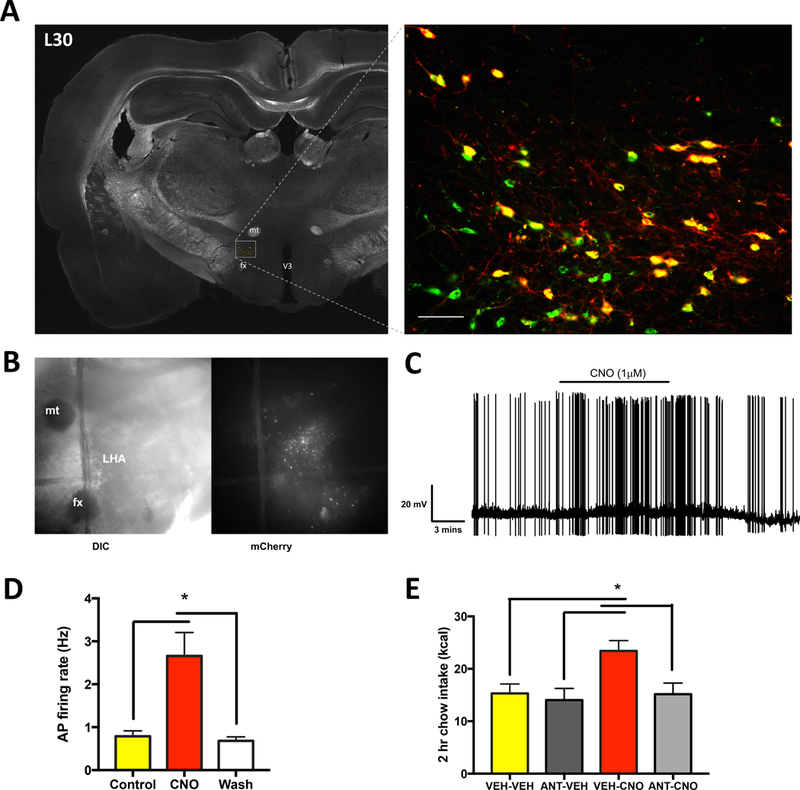
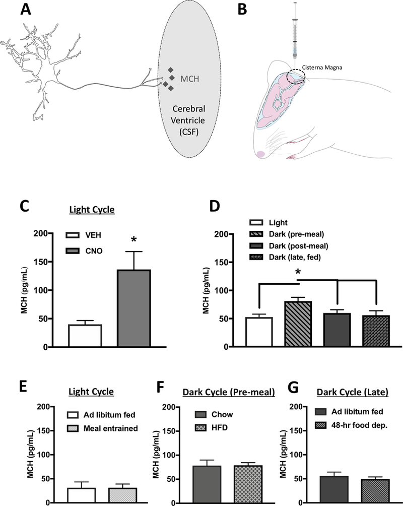
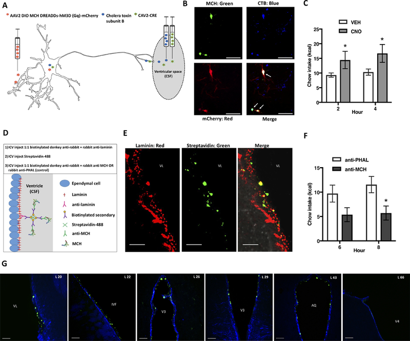
Classical mechanisms through which brain-derived molecules influence behavior include neuronal synaptic communication and neuroendocrine signaling. Here we provide evidence for an alternative neural communication mechanism that is relevant for food intake control involving cerebroventricular volume transmission of the neuropeptide melanin-concentrating hormone (MCH). Results reveal that the cerebral ventricles receive input from approximately one-third of MCH-producing neurons. Moreover, MCH cerebrospinal fluid (CSF) levels increase prior to nocturnal feeding and following chemogenetic activation of MCH-producing neurons. Utilizing a dual viral vector approach, additional results reveal that selective activation of putative CSF-projecting MCH neurons increases food intake. In contrast, food intake was reduced following immunosequestration of MCH endogenously present in CSF, indicating that neuropeptide transmission through the cerebral ventricles is a physiologically relevant signaling pathway for energy balance control. Collectively these results suggest that neural-CSF volume transmission signaling may be a common neurobiological mechanism for the control of fundamental behaviors.

Keywords: CSF; MCH; appetite; cerebrospinal fluid; circadian; feeding; neuroendocrine; obesity; orexin; volume transmission.


Figures

Figure 1:
Figure 1:. MCH-producing neurons terminate in the ependymal layer lining the cerebral ventricles.
Immunofluorescence co-labeling of MCH and synaptophysin at the ventricular ependymal layer supports the possibility of cerebroventricular volume transmission. Red = MCH axons, green = synaptophysin (synaptic vesicle marker), blue = vimentin (ventricular ependymal cell marker). Arrows indicate MCH+ synaptophysin colabeling. Numbers in the upper left in each image preceded by “L” indicate correspondence to closest atlas levels of the rat brain atlas of Swanson (Swanson, 2004). aco = anterior commissure; AQ = cerebral aqueduct; CB = cerebellum; fr = fasciculus retroflexus; LSr = lateral septal nucleus, rostral part; MEex = median eminence; MEin = median eminence, internal lamina; PAG = periaqueductal gray; PCG = pontine central gray; PH = posterior hypothalamic nucleus; PVH, paraventricular hypothalamic nucleus; PVT = paraventricular thalamic nucleus; V3 = third ventricle; V3t = third ventricle, thalamic part; V4 = fourth ventricle; VL = lateral ventricle. Scale bars: white = 10 μm, yellow = 100 μm.
Figure 2:
Figure 2:. Approximately one-third of all MCH neurons are CSF-projecting and these neurons are distinct from neuroendocrine neurons.
(A) Distribution of MCH immunolabeled and CTB retrogradely labeled neurons following lateral ventricular CTB injections; representative immunofluorescence for MCH (green), CTB (red), and colabeled cells (yellow). Numbers in the lower left in each image preceded by “L” indicate correspondence to closest atlas levels of the rat brain atlas of Swanson (Swanson, 2004). (B) Overall 32.7% of MCH-ir somata within the LHA and ZI were also CTB-ir, whereas 43.8 % of CTB-ir somata in the LHA and ZI were MCH-ir. Percentages of colabeling for the ZI and for specific LHA subregions are represented as follows: blue bars indicate the percentage of total (across all brain regions) colocalized MCH-ir + CTB-ir located in each region; red bars indicate the percentage CTB-ir neurons that are colocalized with MCH-ir in each region; green bars indicate the percentage MCH-ir neurons that are colocalized with CTB-ir in each region. (C) Representative images of immunofluorescence for MCH (green), CTB (red) and Fast Blue (FB, blue) in the lateral hypothalamic area. Intravenously injected FB retrogradely labelled neurons with access to the vasculature (putative neuroendocrine neurons). Overall 98.8% of MCH + CTB analyzed were not labeled with FB, with the arrow pointing to an extremely rare instance of a triple labeled cell (MCH + CTB + FB). Abbreviations: d = LHA dorsal region; fx = fornix; jd = LHA juxtadorsomedial region; jp = lateral hypothalamic area (LHA) juxtaparaventricular region; jvd = LHA juxtaventromedial region, dorsal zone; jvv = LHA juxtaventromedial region, ventral zone; m = LHA magnocellular nucleus; mtt = mammillothalamic tract; p = LHA posterior region; pc = LHA parvicellular region; PH = posterior hypothalamic nucleus; PST = preparasubthalamic nucleus; s = LHA suprafornical region; sfa = LHA subfornical region, anterior zone; sfp = LHA subfornical region, posterior zone; TUl = tuberal nucleus, lateral part; vl = LHA ventral region, lateral zone; vm = LHA medial zone, ventral region; ZI = zona incerta. Scale bar = 50 μm.
Figure 3:
Figure 3:. Approximately 40% of hypocretin /orexin-producing neurons project to CSF.
(A) Representative images of cholera toxin subunit B (CTB, red), hypocretin/orexin (H/O, green) and colabeled neurons (yellow) in the LHA. The presence of CTB retrograde labeling in H/O-ir neurons after CTB injection into the lateral ventricle indicates that some H/O neurons project to the CSF. Abbreviations: fx = fornix. Scale bars = 50 μm. (B) Immunofluorescence colabeling of H/O and synaptophysin at the cerebroventricular ependyma supports the possibility of volume transmission into the CSF. Synaptophysin (a synaptic vesicle marker) and H/O colabeling was present at the site of ependymal cells immunolabeled for vimentin (a marker of ependymal cells) at multiple locations within the cerebral ventricles. Red = H/O axons, green = synaptophysin, blue = vimentin. Arrows indicate H/O + synaptophysin colabeling. Numbers upper left in each image preceded by “L” indicate correspondence to closest atlas levels of the rat brain atlas of Swanson (Swanson, 2004). Abbreviations: aco =anterior commissure; AQ = cerebral aqueduct; LSr, lateral septal nucleus, rostral part; VL = lateral ventricle; ME = median eminence; PAG =periaqueductal gray; PH = posterior hypothalamic nucleus; PVH = hypothalamic paraventricular nucleus; PVT = paraventricular thalamic nucleus; V3, third ventricle. Scale bars = 100 μm.
Figure 4:
Figure 4:. While 45% of CSF-projecting MCH neurons co-express GABAergic markers, GABA signaling is unlikely to interact with MCH-driven hyperphagia.
(A) CTB was injected into the lateral ventricle and immunofluorescent staining was performed for MCH (green), CTB (red) and the GABAergic marker GAD67 (blue). Quantification indicated that 14.4% of MCH neurons were both GABAergic (GAD67 immunolabeled) and CSF-signaling (CTB retrogradely labeled following CTB injection into the lateral ventricle), whereas 17.6 % of CTB positive MCH neurons did not contain the GABAergic marker. Thus 45% of putative CSF-signaling MCH neurons were GABAergic, compared to 23.5% of MCH neurons that are putative non-CSF-signaling. Scale bar: 50 μm. (B) CTB was injected into the lateral ventricle and immunofluorescent staining was performed for MCH (red) and CTB (green), followed by fluorescence in situ hybridization for the glutamatergic marker vGLUT2 (purple). Quantification indicated that there was no colocalization of vGLUT2 in CTB positive MCH-ir neurons (Scale bar: 100 μm), nor was there colocalization of vGLUT1 or vGLUT3 (data not shown). (C) A dose response for the effects of ICV injections of the GABA-A receptor agonist muscimol indicated a significant effect of drug where 75 ng significantly elevated food intake while 25 ng was ineffective (n=12). (D) ICV injection of a sub-effective dose of muscimol (20 ng) did not potentiate the feeding effects of a sub-effective dose of MCH (0.5 μg)(n=12). (E) ICV injection of a sub-effective dose of Baclofen (0.1 nmol) did not potentiate the feeding effects of a sub-effective dose of MCH (0.5 μg)(n=19) (*P<.05).
Figure 5:
Figure 5:. Viral transduction of Designer Receptors Exclusively Activated by Designer Drugs (DREADDs; hM3Gq) is a valid method of inducing selective and functionally-relevant activation of MCH neurons.
(A) Representative images of the mCherry reporter gene present in MCH neurons, indicating successful infection of the DREADDS AAV selective in MCH neurons. MCH: Green; mCherry: Red; Colocalization: Yellow. Scale bar: 50 μm. (B) Representative differential interference contrast (DIC) and fluorescence microscopy images of hypothalamic slice preparations. (C) Representative trace of electrophysiological recordings from mCherry positive DREADDs-expressing MCH neurons before, during, and after clozapine-N-oxide (CNO; the DREADDs ligand) application. (D) Rats were bilaterally injected in the lateral hypothalamic area and zona incerta with an AAV driving hM3Gq DREADDS expression under the control of a promoter for MCH (AAV2-rMCHp-hM3D(Gq)-mCherry). Data from electrophysiological recordings showing that CNO significantly increased the firing rate of mCherry positive DREADDs-expressing MCH neurons (n=8). (E) CNO (18 mmoles) was used to pharmacologically activate MCH neurons, which resulted in increased feeding that was blocked by pretreatment with the MCH 1R antagonist H6408 (ANT; 10μg)(n=7,8/group) (*P<.05). All data are represented as means ± SEM. mt, mammillothalamic tract; fx, fornix; V3, third ventricle.
Figure 6:
Figure 6:. MCH levels in the CSF are increased by DREADDs-mediated activation of MCH neurons and prior to nocturnal feeding.
(A) A hypothetical model whereby MCH is transmitted into the CSF through axon terminals of ventricular-contacting terminals from MCH neurons. (B) Cartoon demonstrating the method of CSF extraction from the cisterna magna of an anesthetized rat. (C) MCH levels were elevated in CSF following MCH DREADDs activation (n=6,7). (D) Under physiological conditions, MCH levels in CSF are elevated during the early dark cycle prior to food consumption compared to during the light cycle and dark cycle postprandially (n=6–8). (E) There were no differences in CSF MCH levels prior to light cycle feeding in meal entrained animals compared to ad libitum fed controls (n=7/group). (F) Five days of exposure to a palatable high-fat diet had no effect on pre-prandial CSF MCH levels during the early dark cycle compared with chow-fed animals (n=6/group). (G) 48 hours of food deprivation had no effect on CSF MCH levels during the late dark cycle compared with ad libitum chow-fed controls. (n=5–6/group) (*P<.05). Data are means ± SEM.
Figure 7:
Figure 7:. Selective activation of ventricular-projecting MCH neurons increases feeding, whereas feeding is reduced by immunosequestration of MCH endogenously present in the CSF.
(A) To selectively activate ventricular projecting MCH neurons, we utilized a dual virus strategy 1) the retrograde canine adenovirus type 2 containing cre recombinase (CAV2-CRE) was injected into the lateral ventricle, and 2) a cre-dependent MCH-DREADDs AAV serotype 2 (AAV2-DIO-MCH DREADDs-hM3D(Gq)-mCherry was injected into the LHA and ZI. (B) Epifluorescence image showing mCherry labeling (red) colocalized with MCH immunolabeling (green), and cells retrogradely labeled by the retrograde tracer cholera toxin subunit B (CTB; blue), following lateral ventricle CTB injection. (C) Activation of ventricular projecting MCH neurons by lateral ventricular injections of CNO increases food intake (n=7). (D) A cartoon depicting the strategy and steps of an immunosequesteration approach designed to reduce the bioavailability of endogenous CSF MCH. (E) A representative image showing immunofluorescence labeling of laminin (red), streptavidin-Alexa Fluor 488 (green) and their colocalization (yellow) at the ventricular ependymal layer (indicating that anti-MCH immunoglobulins were localized with laminin-ir at the ventricular ependyma). (F) Results from the immuno-sequestration experiment indicated that under normal physiological conditions a reduction in the bioavailability of CSF MCH significantly reduces food intake (n=8,9). (G) Representative immunofluorescence images showing the rostral-caudal spread of streptavidin-Alexa Fluor 488 (green) localized at the ventricular ependyma (shown with vimentin immunofluorescence in blue), indicating extensive rostral caudal CSF MCH immunosequestration. The numbers in the upper left in each image preceded by “L” indicate correspondence to closest atlas levels of the rat brain atlas of Swanson (Swanson, 2004) (*P<.05). Data are means ± SEM. Scale bars in B, E = 50 μm, scale bars in F = 100μm. Abbreviations: AL = atlas level; AQ = cerebral aqueduct; IVF = Intraventricular foramen; V3 = third ventricle; V4 = fourth ventricle; VL = lateral ventricle.

Comment in

References

    1. Agnati LF, Fuxe K, Zoli M, Ozini I, Toffano G, and Ferraguti F (1986). A correlation analysis of the regional distribution of central enkephalin and beta-endorphin immunoreactive terminals and of opiate receptors in adult and old male rats. Evidence for the existence of two main types of communication in the central nervous system: the volume transmission and the wiring transmission. Acta physiologica Scandinavica 128, 201–207. - PubMed
    1. Balland E, Dam J, Langlet F, Caron E, Steculorum S, Messina A, Rasika S, Falluel-Morel A, Anouar Y, Dehouck B, et al. (2014). Hypothalamic tanycytes are an ERK-gated conduit for leptin into the brain. Cell metabolism 19, 293–301. - PMC - PubMed
    1. Barson JR, Morganstern I, and Leibowitz SF (2013). Complementary roles of orexin and melanin-concentrating hormone in feeding behavior. Int J Endocrinol 2013, 983964. - PMC - PubMed
    1. Bittencourt JC, Presse F, Arias C, Peto C, Vaughan J, Nahon JL, Vale W, and Sawchenko PE (1992). The melanin-concentrating hormone system of the rat brain: an immuno- and hybridization histochemical characterization. J Comp Neurol 319, 218–245. - PubMed
    1. Bjorefeldt A, Andreasson U, Daborg J, Riebe I, Wasling P, Zetterberg H, and Hanse E (2015). Human cerebrospinal fluid increases the excitability of pyramidal neurons in the in vitro brain slice. The Journal of physiology 593, 231–243. - PMC - PubMed