Renal Vein Thrombosis as Presentation of Non-M3 Acute Myeloid Leukemia in an Adult Patient
- PMID: 29861566
- PMCID: PMC5952454
- DOI: 10.4103/ijn.IJN_54_17
Renal Vein Thrombosis as Presentation of Non-M3 Acute Myeloid Leukemia in an Adult Patient
Abstract
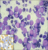
A 46-year-old male presented with left flank pain and was found to have left nephromegaly with renal vein and inferior vena cava (IVC) thrombus. On hematological evaluation, he had leukocytosis and thrombocytopenia. Further evaluation revealed acute myeloid leukemia (AML). Following initial cytoreductive therapy and supportive care for hyperleukocytosis, he underwent left simple nephrectomy with IVC thrombectomy. Postoperatively, he developed massive thrombosis of infrahepatic IVC with renal failure. Renal venous thrombosis as a rare presentation of AML in adults with leukemic hyperleukocytosis has not been reported. In the absence of clear guidelines, early diagnosis and management are desirable.
Keywords: Acute myeloid leukemia; renal vein thrombus; thrombectomy.
Conflict of interest statement
There are no conflicts of interest.
Figures


References
-
- Creutzig U, Ritter J, Budde M, Sutor A, Schellong G. Early deaths due to hemorrhage and leukostasis in childhood acute myelogenous leukemia. Associations with hyperleukocytosis and acute monocytic leukemia. Cancer. 1987;60:3071–9. - PubMed
-
- Murray JC, Dorfman SR, Brandt ML, Dreyer ZE. Renal venous thrombosis complicating acute myeloid leukemia with hyperleukocytosis. J Pediatr Hematol Oncol. 1996;18:327–30. - PubMed
-
- De Stefano V, Sorà F, Rossi E, Chiusolo P, Laurenti L, Fianchi L, et al. The risk of thrombosis in patients with acute leukemia: Occurrence of thrombosis at diagnosis and during treatment. J Thromb Haemost. 2005;3:1985–92. - PubMed
-
- Kwaan HC, Vicuna B. Incidence and pathogenesis of thrombosis in hematologic malignancies. Semin Thromb Hemost. 2007;33:303–12. - PubMed
Publication types
LinkOut - more resources
Full Text Sources
Other Literature Sources

