Review
. 2018 May 14:2018:8430123.
doi: 10.1155/2018/8430123. eCollection 2018.

Tumor Necrosis Factor and Interleukin-1 β Modulate Synaptic Plasticity during Neuroinflammation

Affiliations
Review

Tumor Necrosis Factor and Interleukin-1 β Modulate Synaptic Plasticity during Neuroinflammation

Francesca Romana Rizzo et al. Neural Plast. .

Abstract

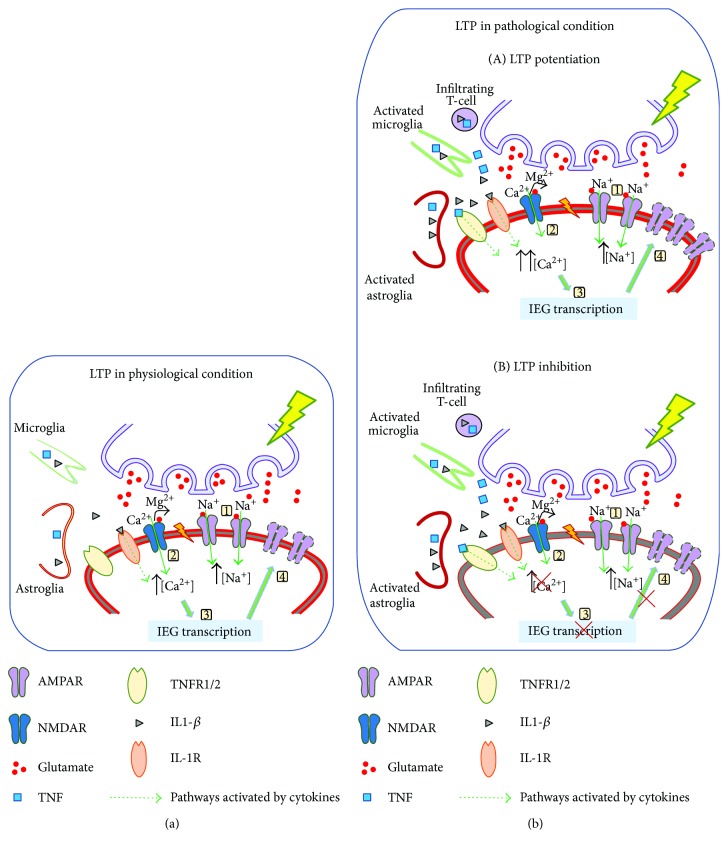
Cytokines are constitutively released in the healthy brain by resident myeloid cells to keep proper synaptic plasticity, either in the form of Hebbian synaptic plasticity or of homeostatic plasticity. However, when cytokines dramatically increase, establishing a status of neuroinflammation, the synaptic action of such molecules remarkably interferes with brain circuits of learning and cognition and contributes to excitotoxicity and neurodegeneration. Among others, interleukin-1β (IL-1β) and tumor necrosis factor (TNF) are the best studied proinflammatory cytokines in both physiological and pathological conditions and have been invariably associated with long-term potentiation (LTP) (Hebbian synaptic plasticity) and synaptic scaling (homeostatic plasticity), respectively. Multiple sclerosis (MS) is the prototypical neuroinflammatory disease, in which inflammation triggers excitotoxic mechanisms contributing to neurodegeneration. IL-β and TNF are increased in the brain of MS patients and contribute to induce the changes in synaptic plasticity occurring in MS patients and its animal model, the experimental autoimmune encephalomyelitis (EAE). This review will introduce and discuss current evidence of the role of IL-1β and TNF in the regulation of synaptic strength at both physiological and pathological levels, in particular speculating on their involvement in the synaptic plasticity changes observed in the EAE brain.


Figures

Figure 1
Figure 1
LTP regulation in physiological and pathological states. The triggering of LTP implies coincident pre- and postsynaptic neuron activation. Strong glutamate release from presynaptic terminal promotes membrane depolarization mediated by Na+ influx through AMPARs (1), which in turn activates NMDARs by means of Mg2+ expulsion from NMDAR pore, thus allowing Ca2+ influx (2). Next, the increase of intracellular Ca2+ concentration activates a cascade of events involving several molecular players and leads to the induction of IEGs (3), such as Arc/Arg, necessary for structural (increased stability and size of dendritic spines) and functional changes of the PSD and the synthesis and insertion of AMPARs in membrane (4). Physiological levels of IL-1β released by both microglia and astroglia contribute to LTP phenomenon (a). During neuroinflammatory disorders (b), activated resident (microglia and astroglia) and infiltrating T-cells strongly release TNF and IL-1β, thus generating two possible outcomes of synaptic changes (A, B). As illustrated in the figure, LTP can be either potentiated or prevented through the action of TNF and IL-1β interfering with the pathways controlling the molecular and structural synaptic changes occurring during LTP.
Figure 2
Figure 2
Synaptic upscaling in response to physiological and pathological stimuli. (a) In a physiological state, during basal synaptic activity, AMPARs undergo constant cycles of membrane insertion and removal on postsynaptic neuron. When the synaptic strength driven by the presynaptic terminal is reduced, TNF, released by astroglia, activates a molecular mechanism leading to transient improved insertion of AMPARs on postsynaptic membrane. (b) During acute or chronic neuroinflammation, TNF, massively released by activated microglia and astroglia as well as infiltrating T-cells, indefinitely upregulates the mechanism of membrane AMPAR insertion. In parallel, inflammation affects physiological mechanisms of glutamate clearance at synaptic cleft. This together with enhanced glutamate release from glial cells over activates AMPARs, thus contributing to induce excitotoxic mechanisms and synaptic loss.

Similar articles

Cited by

References

    1. McAfoose J., Baune B. T. Evidence for a cytokine model of cognitive function. 2009;33(3):355–366. doi: 10.1016/j.neubiorev.2008.10.005. - DOI - PubMed
    1. Schwartz M., Deczkowska A. Neurological disease as a failure of brain–immune crosstalk: the multiple faces of neuroinflammation. 2016;37(10):668–679. doi: 10.1016/j.it.2016.08.001. - DOI - PubMed
    1. Di Filippo M., Sarchielli P., Picconi B., Calabresi P. Neuroinflammation and synaptic plasticity: theoretical basis for a novel, immune-centred, therapeutic approach to neurological disorders. 2008;29(8):402–412. doi: 10.1016/j.tips.2008.06.005. - DOI - PubMed
    1. Yirmiya R., Goshen I. Immune modulation of learning, memory, neural plasticity and neurogenesis. 2011;25(2):181–213. doi: 10.1016/j.bbi.2010.10.015. - DOI - PubMed
    1. Yang G., Parkhurst C. N., Hayes S., Gan W.-B. Peripheral elevation of TNF-α leads to early synaptic abnormalities in the mouse somatosensory cortex in experimental autoimmune encephalomyelitis. 2013;110(25):10306–10311. doi: 10.1073/pnas.1222895110. - DOI - PMC - PubMed

Publication types

MeSH terms

Substances