

Multifactorial Analysis of Mortality in Screening Detected Lung Cancer
- PMID: 29861726
- PMCID: PMC5976935
- DOI: 10.1155/2018/1296246
Multifactorial Analysis of Mortality in Screening Detected Lung Cancer
Abstract
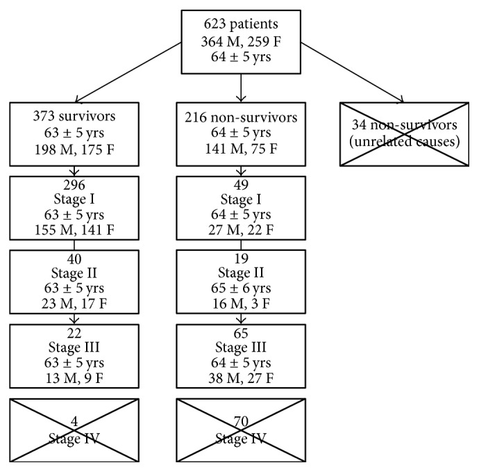
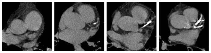
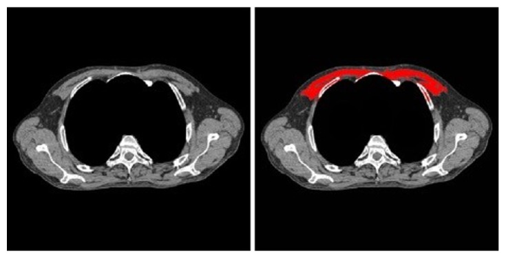
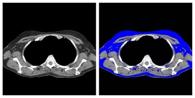
We hypothesized that severity of coronary artery calcification (CAC), emphysema, muscle mass, and fat attenuation can help predict mortality in patients with lung cancer participating in the National Lung Screening Trial (NLST). Following regulatory approval from the Cancer Data Access System (CDAS), all patients diagnosed with lung cancer at the time of the screening study were identified. These subjects were classified into two groups: survivors and nonsurvivors at the conclusion of the NLST trial. These groups were matched based on their age, gender, body mass index (BMI), smoking history, lung cancer stage, and survival time. CAC, emphysema, muscle mass, and subcutaneous fat attenuation were quantified on baseline low-dose chest CT (LDCT) for all patients in both groups. Nonsurvivor group had significantly greater CAC, decreased muscle mass, and higher fat attenuation compared to the survivor group (p < 0.01). No significant difference in severity of emphysema was noted between the two groups (p > 0.1). We thus conclude that it is possible to create a quantitative prediction model for lung cancer mortality for subjects with lung cancer detected on screening low-dose CT (LDCT).
Figures
Similar articles
-
AI Body Composition in Lung Cancer Screening: Added Value Beyond Lung Cancer Detection.Radiology. 2023 Jul;308(1):e222937. doi: 10.1148/radiol.222937. Radiology. 2023. PMID: 37489991 Free PMC article.
-
The effect of direct referral for fast CT scan in early lung cancer detection in general practice. A clinical, cluster-randomised trial.Dan Med J. 2015 Mar;62(3):B5027. Dan Med J. 2015. PMID: 25748876 Clinical Trial.
-
Decreased cardiovascular mortality in the ITALUNG lung cancer screening trial: Analysis of underlying factors.Lung Cancer. 2019 Dec;138:72-78. doi: 10.1016/j.lungcan.2019.10.006. Epub 2019 Oct 15. Lung Cancer. 2019. PMID: 31654837
-
Extracoronary Thoracic and Coronary Artery Calcifications on Chest CT for Lung Cancer Screening: Association with Established Cardiovascular Risk Factors - The "CT-Risk" Trial.Acad Radiol. 2015 Jul;22(7):880-9. doi: 10.1016/j.acra.2015.03.005. Epub 2015 May 7. Acad Radiol. 2015. PMID: 25957500 Review.
-
To Screen or not to Screen: Low Dose Computed Tomography in Comparison to Chest Radiography or Usual Care in Reducing Morbidity and Mortality from Lung Cancer.Cureus. 2016 Apr 27;8(4):e589. doi: 10.7759/cureus.589. Cureus. 2016. PMID: 27375974 Free PMC article. Review.
Cited by
-
Preoperative pectoralis muscle index predicts distant metastasis-free survival in non-small cell lung cancer patients: a retrospective study.BMC Med Imaging. 2025 Aug 19;25(1):335. doi: 10.1186/s12880-025-01873-0. BMC Med Imaging. 2025. PMID: 40830848 Free PMC article.
-
Quantitative Pectoralis Muscle Area is Associated with the Development of Lung Cancer in a Large Lung Cancer Screening Cohort.Lung. 2020 Oct;198(5):847-853. doi: 10.1007/s00408-020-00388-5. Epub 2020 Sep 5. Lung. 2020. PMID: 32889594 Free PMC article.
-
Artificial intelligence-based vessel suppression for detection of sub-solid nodules in lung cancer screening computed tomography.Quant Imaging Med Surg. 2021 Apr;11(4):1134-1143. doi: 10.21037/qims-20-630. Quant Imaging Med Surg. 2021. PMID: 33816155 Free PMC article.
-
Can CT radiomic analysis in NSCLC predict histology and EGFR mutation status?Medicine (Baltimore). 2019 Jan;98(1):e13963. doi: 10.1097/MD.0000000000013963. Medicine (Baltimore). 2019. PMID: 30608433 Free PMC article.
-
AI Body Composition in Lung Cancer Screening: Added Value Beyond Lung Cancer Detection.Radiology. 2023 Jul;308(1):e222937. doi: 10.1148/radiol.222937. Radiology. 2023. PMID: 37489991 Free PMC article.
References
-
- International Early Lung Cancer Action Program Investigators, Henschke C. I., Yankelevitz D. F., et al. Survival of patients with stage I lung cancer detected on CT screening. 2006;355:1763–1771. - PubMed
LinkOut - more resources
Full Text Sources
Other Literature Sources
