A Review on Biomechanics of Anterior Cruciate Ligament and Materials for Reconstruction
- PMID: 29861784
- PMCID: PMC5971278
- DOI: 10.1155/2018/4657824
A Review on Biomechanics of Anterior Cruciate Ligament and Materials for Reconstruction
Abstract
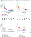
The anterior cruciate ligament is one of the six ligaments in the human knee joint that provides stability during articulations. It is relatively prone to acute and chronic injuries as compared to other ligaments. Repair and self-healing of an injured anterior cruciate ligament are time-consuming processes. For personnel resuming an active sports life, surgical repair or replacement is essential. Untreated anterior cruciate ligament tear results frequently in osteoarthritis. Therefore, understanding of the biomechanics of injury and properties of the native ligament is crucial. An abridged summary of the prominent literature with a focus on key topics on kinematics and kinetics of the knee joint and various loads acting on the anterior cruciate ligament as a function of flexion angle is presented here with an emphasis on the gaps. Briefly, we also review mechanical characterization composition and anatomy of the anterior cruciate ligament as well as graft materials used for replacement/reconstruction surgeries. The key conclusions of this review are as follows: (a) the highest shear forces on the anterior cruciate ligament occur during hyperextension/low flexion angles of the knee joint; (b) the characterization of the anterior cruciate ligament at variable strain rates is critical to model a viscoelastic behavior; however, studies on human anterior cruciate ligament on variable strain rates are yet to be reported; (c) a significant disparity on maximum stress/strain pattern of the anterior cruciate ligament was observed in the earlier works; (d) nearly all synthetic grafts have been recalled from the market; and (e) bridge-enhanced repair developed by Murray is a promising technique for anterior cruciate ligament reconstruction, currently in clinical trials. It is important to note that full extension of the knee is not feasible in the case of most animals and hence the loading pattern of human ACL is different from animal models. Many of the published reviews on the ACL focus largely on animal ACL than human ACL. Further, this review article summarizes the issues with autografts and synthetic grafts used so far. Autografts (patellar tendon and hamstring tendon) remains the gold standard as nearly all synthetic grafts introduced for clinical use have been withdrawn from the market. The mechanical strength during the ligamentization of autografts is also highlighted in this work.
Figures


References
-
- Tortora G. J., Derrickson B. H. 12th. Hoboken, NJ, USA: John Wiley & Sons Inc; 2009.
-
- Boden B. P., Dean G. S., Feagin J. A., Jr., Garrett W. E., Jr. Mechanisms of anterior cruciate ligament injury. 2000;23(6):573–578. - PubMed
-
- McNair P. J., Marshall R. N., Matheson J. A. Important features associated with acute anterior cruciate ligament injury. 1990;103(901):537–539. - PubMed
Publication types
LinkOut - more resources
Full Text Sources
Other Literature Sources

