

Case Report: "Incognito" proteus syndrome
- PMID: 29862018
- PMCID: PMC5843845
- DOI: 10.12688/f1000research.13993.1
Case Report: "Incognito" proteus syndrome
Abstract
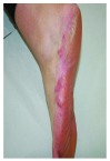
Proteus syndrome (PS) is a postnatal mosaic overgrowth disorder, progressive and disfiguring. It is clinically diagnosed according to the criteria reported by Biesecker et al. We describe the case of a 49-year-old woman who presented with a 10-year history of pauci-symptomatic infiltrating plaque lesions on the sole and lateral margin of the left foot, which had been diagnosed as a keloid. The patient had a positive history for advanced melanoma and a series of subtle clinical signs, such as asymmetric face, scoliosis, multiple lipomas on the trunk, linear verrucous epidermal nevi, and hyperpigmented macules with a mosaic distribution. Even if the clinical presentation was elusive, she had enough criteria to be diagnosed with PS. This case describes the first evidence, to the best of our knowledge, of pauci-symptomatic PS in adulthood, reports its rare association with advanced melanoma, and illustrates the importance of even minor cutaneous clinical signs, especially when atypical, in formulating the diagnosis of a complex cutaneous condition such as this.
Keywords: cerebriform; diagnosis; elusive; hidden; keloid; proteus syndrome.
Conflict of interest statement
No competing interests were disclosed.
Figures


Similar articles
-
Cerebriform fibrous proliferation vs. proteus syndrome.Ann Plast Surg. 2001 Dec;47(6):669-72. doi: 10.1097/00000637-200112000-00016. Ann Plast Surg. 2001. PMID: 11756840
-
Proteus syndrome: A case report and review of the literature.Mol Clin Oncol. 2017 Mar;6(3):381-383. doi: 10.3892/mco.2017.1140. Epub 2017 Jan 23. Mol Clin Oncol. 2017. PMID: 28451417 Free PMC article.
-
Early Recognition of Proteus Syndrome.Pediatr Dermatol. 2016 Sep;33(5):e306-10. doi: 10.1111/pde.12900. Epub 2016 Jul 4. Pediatr Dermatol. 2016. PMID: 27378680
-
Proteus syndrome: three case reports with a review of the literature.Fetal Pediatr Pathol. 2012 Jun;31(3):145-53. doi: 10.3109/15513815.2012.656830. Epub 2012 Mar 13. Fetal Pediatr Pathol. 2012. PMID: 22413928 Review.
-
Proteus syndrome: a natural clinical course of Proteus syndrome.Yonsei Med J. 2002 Apr;43(2):259-66. doi: 10.3349/ymj.2002.43.2.259. Yonsei Med J. 2002. PMID: 11971221 Review.
Cited by
-
Proteus syndrome of the foot: A case report and literature review.Exp Ther Med. 2020 Sep;20(3):2716-2720. doi: 10.3892/etm.2020.8986. Epub 2020 Jul 10. Exp Ther Med. 2020. PMID: 32765766 Free PMC article.
-
Intraspinal lipomatous neurofibroma in a child with atypical Proteus syndrome.Childs Nerv Syst. 2024 Dec;40(12):3895-3900. doi: 10.1007/s00381-024-06652-w. Epub 2024 Oct 21. Childs Nerv Syst. 2024. PMID: 39432065 No abstract available.
References
-
- Cohen MM, Jr, Hayden PW: A newly recognized hamartomatous syndrome. Birth Defects. 1979;15(5B):291–296. - PubMed
-
- Biesecker LG, Sapp JC: Proteus Syndrome.2012.
LinkOut - more resources
Full Text Sources
Other Literature Sources