Review
. 2018 May 29:4:12.
doi: 10.1186/s40842-018-0062-7. eCollection 2018.

The challenges of diagnosing osteoporosis and the limitations of currently available tools

Affiliations
Review

The challenges of diagnosing osteoporosis and the limitations of currently available tools

Palak Choksi et al. Clin Diabetes Endocrinol. .

Abstract

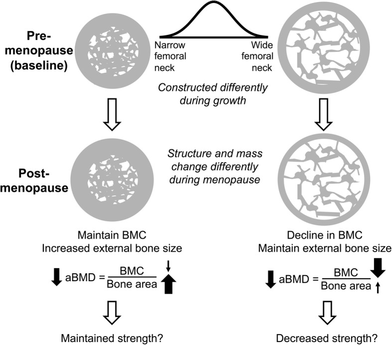
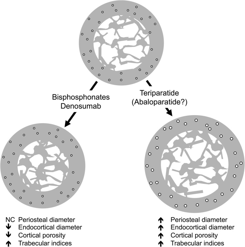
Dual-energy X-ray absorptiometry (DXA) was the first imaging tool widely utilized by clinicians to assess fracture risk, especially in postmenopausal women. The development of DXA nearly coincided with the availability of effective osteoporosis medications. Although osteoporosis in adults is diagnosed based on a T-score equal to or below - 2.5 SD, most individuals who sustain fragility fractures are above this arbitrary cutoff. This incongruity poses a challenge to clinicians to identify patients who may benefit from osteoporosis treatments. DXA scanners generate 2 dimensional images of complex 3 dimensional structures, and report bone density as the quotient of the bone mineral content divided by the bone area. An obvious pitfall of this method is that a larger bone will convey superior strength, but may in fact have the same bone density as a smaller bone. Other imaging modalities are available such as peripheral quantitative CT, but are largely research tools. Current osteoporosis medications increase bone density and reduce fracture risk but the mechanisms of these actions vary. Anti-resorptive medications (bisphosphonates and denosumab) primarily increase endocortical bone by bolstering mineralization of endosteal resorption pits and thereby increase cortical thickness and reduce cortical porosity. Anabolic medications (teriparatide and abaloparatide) increase the periosteal and endosteal perimeters without large changes in cortical thickness resulting in a larger more structurally sound bone. Because of the differences in the mechanisms of the various drugs, there are likely benefits of selecting a treatment based on a patient's unique bone structure and pattern of bone loss. This review retreats to basic principles in order to advance clinical management of fragility fractures by examining how skeletal biomechanics, size, shape, and ultra-structural properties are the ultimate predictors of bone strength. Accurate measurement of these skeletal parameters through the development of better imaging scanners is critical to advancing fracture risk assessment and informing clinicians on the best treatment strategy. With this information, a "treat to target" approach could be employed to tailor current and future therapies to each patient's unique skeletal characteristics.

Keywords: Bisphosphonates; Denosumab; Dual X-ray absorptiometry; Osteoporosis; Peripheral quantitative computed tomography; Romosozumab; Skeletal biomechanics; Skeletal fracture; Teriparatide.


Conflict of interest statement

Not applicable.The authors declare that they have no competing interests.Springer Nature remains neutral with regard to jurisdictional claims in published maps and institutional affiliations.

Figures

Fig. 1
Fig. 1
Structural characteristics of bone. Bone is comprised of a dense cortical shell that surrounds a spongy trabecular bone network. The periosteal diameter combined with the endosteal diameter determines cortical thickness. The size of bone along with cortical thickness and porosity significantly contribute to bone strength. The inner trabecular compartment contains a network of plates and rods that also contribute to bone strength
Fig. 2
Fig. 2
Determinants of bone strength. Bone strength is a composite summation of numerous skeletal characteristics. The size of bone increases with age and with puberty. Ultimate bone size also has a large genetic contribution. Genetics and habitual loading determine bone shape. The architecture of bone is a complex interplay among many structural components. Cortical diameter, thickness and porosity contribute to cortical strength. The number, thickness, and the connectivity of plates and rods determine trabecular bone strength. Bone composition is difficult to measure non-invasively. The degree of collagen crosslinking and the density of collagen contribute to bone matrix strength. Newly formed protein matrix subsequently becomes mineralized and how the hydroxyapatite crystals are arranged within the matrix and the degree of mineralization contribute to bone hardness and strength
Fig. 3
Fig. 3
Strategic arrangement of cortical and trabecular bone. The proximal femur experiences forces in different directions. a The critical aspects of femoral neck strength superimposed onto a hip DXA scan image. b With standing, the femoral neck experiences compress forces on the inferior surface and tensile forces on the superior surface. Compressive loads are reinforced with a compressive arcade composed of a thickened inferior cortex and an additional trabecular network. The tensile arcade is reinforced with a network of trabecular bone. These reinforcements are combined with lateral and medial cortices that provide additional reinforcements against side-to-side forces. NanoCT images were taken at 27 μm resolution using a phoenix nanotom-s (GE Sensing and Inspection Technologies, GmbH, Wunstorf, Germany)
Fig. 4
Fig. 4
Areal BMD as determined by DXA declines with aging for different reasons. With aging, women with smaller femoral necks tend to increase bone area through an increase in cortical thickness by an increase in periosteal and endosteal bone formation. Since BMD may only decrease slightly but bone area increases more, the result is lower areal BMD as measured by DXA despite likely having little change in bone strength. In the case of women with larger femoral necks, the endosteal cortex undergoes excessive resorption without periosteal expansion resulting in a thinner cortex. The result is a lower BMC without significant change in bone area. The DXA areal BMD decreases and may result in a bone with less strength. Adapted from Jepsen, et al. JBMR 2017 [32]
Fig. 5
Fig. 5
Structural changes in bone with osteoporosis medications. The anti-resorptive medications (bisphosphonates and denosumab) and anabolic medications (teriparatide and likely abaloparatide) produce very different structural changes in bone. Although both classes increase trabecular bone, their effects on cortical bone are different. Bisphosphonates and denosumab do not expand periosteal bone but do decrease the endosteal diameter by an increase in endosteal bone volume. Anti-resorptives also reduce cortical porosity. Anabolic agents lead to an increase in periosteal bone with a simultaneous increase in endosteal bone resorption resulting in a bone without a large change in cortical thickness. At the same time, anabolic agents increase cortical porosity. Despite the increase in cortical porosity, the larger bone has increased strength. NC = no change

References

    1. What is Osteoporosis and What Causes It? Arlington: National Osteoporosis Foundation. https://www.nof.org/patients/what-is-osteoporosis/.
    1. Wahner HW, Dunn WL, Brown ML, Morin RL, Riggs BL. Comparison of dual-energy x-ray absorptiometry and dual photon absorptiometry for bone mineral measurements of the lumbar spine. Mayo Clin Proc. 1988;63:1075–1084. doi: 10.1016/S0025-6196(12)65502-5. - DOI - PubMed
    1. Kanis JA. Assessment of fracture risk and its application to screening for postmenopausal osteoporosis: synopsis of a WHO report. WHO study group. Osteoporos Int. 1994;4:368–381. doi: 10.1007/BF01622200. - DOI - PubMed
    1. Sanders KM, Nicholson GC, Watts JJ, Pasco JA, Henry MJ, Kotowicz MA, Seeman E. Half the burden of fragility fractures in the community occur in women without osteoporosis. When is fracture prevention cost-effective? Bone. 2006;38:694–700. doi: 10.1016/j.bone.2005.06.004. - DOI - PubMed
    1. Kanis JA, McCloskey EV, Johansson H, Oden A, Melton LJ, 3rd, Khaltaev N. A reference standard for the description of osteoporosis. Bone. 2008;42:467–475. doi: 10.1016/j.bone.2007.11.001. - DOI - PubMed