

Case Reports
doi: 10.1155/2018/1301072.
eCollection 2018.
Case Report of Hydatid Cyst in the Pulmonary Artery Uncommon Presentation: CT and MRI Findings
Affiliations
- PMID: 29862110
- PMCID: PMC5976941
- DOI: 10.1155/2018/1301072
Item in Clipboard
Case Reports
Case Report of Hydatid Cyst in the Pulmonary Artery Uncommon Presentation: CT and MRI Findings
Case Rep Radiol.
.
Abstract
Background: Hydatid cysts can be found in any organ. In adults, the liver and lungs are the most common locations; hydatid cysts in the pulmonary artery are rare.
Clinical case: We present the case of an 86-year-old female with a history of hepatic hydatid cyst since 2012, who presented with complaints of chronic productive cough, yellowish-green sputum, and dyspnea. CT and MRI showed multiseptate hydatid cysts in the right pulmonary artery.
Figures

(a-b) CT axial images show pulmonary artery with multiseptate hydatid cysts (red arrows), seen in the right main pulmonary artery and the lower lobar branches of the right pulmonary artery, along with other parenchymal cysts (blue arrows).

(a-b) CT coronal images show right main pulmonary artery hydatid cysts (red arrows) along with a cyst of the right hepatic lobe (blue arrows).

CT axial image shows a large cyst in the right lower lobe representing parenchymal hydatid cyst (yellow arrow); there is also a large cyst with air seen in the left lower lobe representing hydatid cyst in the left lung (green arrow).

(a-b) Axial T1 and T2 weighted images show hydatid cyst (red arrows) in the right main pulmonary artery and small pleural effusions on the left side (green arrows).
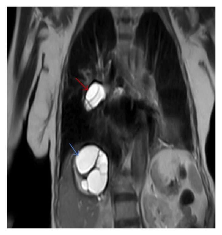
A coronal T2 weighted image shows a high signal intensity cystic lesion (red arrow) within the right main pulmonary artery with multiseptate high intensity cystic lesion on the right hepatic lobe (blue arrow).

Axial T2 weighted image shows a large cyst in the right lower lobe representing parenchymal hydatid cyst (blue arrow); there is also a large cyst with air seen in the left lower lobe representing hydatid cyst in the left lung (green arrow).
Similar articles
-
More than 200 hydatid cysts in the lung with a large number of daughter cysts: A rare case report.Int J Surg Case Rep. 2024 Jan;114:109169. doi: 10.1016/j.ijscr.2023.109169. Epub 2023 Dec 17. Int J Surg Case Rep. 2024. PMID: 38134619 Free PMC article.
-
Hydatid Cyst in the Pulmonary Artery: An Uncommon Localization.Heart Surg Forum. 2004 Jan 1;7(1):13-15. doi: 10.1532/hsf.954. Heart Surg Forum. 2004. PMID: 14980840
-
Concurrent pulmonary and hepatic hydatid cysts managed with single stage surgery.Radiol Case Rep. 2019 Aug 20;14(10):1306-1310. doi: 10.1016/j.radcr.2019.08.006. eCollection 2019 Oct. Radiol Case Rep. 2019. PMID: 31467627 Free PMC article.
-
Hydatid Pulmonary Embolism: A Case Report and Literature Review.Am J Case Rep. 2021 Nov 30;22:e934157. doi: 10.12659/AJCR.934157. Am J Case Rep. 2021. PMID: 34845180 Free PMC article. Review.
-
Rare reason for pulmonary embolism: one case of pulmonary hydatid cyst and review of the literature.J Thromb Thrombolysis. 2015 Jul;40(1):126-9. doi: 10.1007/s11239-014-1147-5. J Thromb Thrombolysis. 2015. PMID: 25359624 Review.
Cited by
-
Primary isolated extraluminal hydatid cyst of left pulmonary artery.Int J Surg Case Rep. 2023 May;106:108211. doi: 10.1016/j.ijscr.2023.108211. Epub 2023 Apr 15. Int J Surg Case Rep. 2023. PMID: 37113706 Free PMC article.
-
The spectrum of imaging findings in pulmonary hydatid disease and the additive value of T2-weighted magnetic resonance imaging in its diagnosis.Pol J Radiol. 2021 Jan 20;86:e53-e63. doi: 10.5114/pjr.2021.103237. eCollection 2021. Pol J Radiol. 2021. PMID: 33708273 Free PMC article.
References
-
- Bakir I., Enc Y., Cicek S. Hydatid cyst in the pulmonary artery: an uncommon localization. 2003;7(1):13–15. - PubMed
Publication types
LinkOut - more resources
Full Text Sources
Other Literature Sources
