. 2018 May 13:2018:4120471.
doi: 10.1155/2018/4120471. eCollection 2018.

Effects of Polylactide Copolymer Implants and Platelet-Rich Plasma on Bone Regeneration within a Large Calvarial Defect in Sheep

Affiliations

Effects of Polylactide Copolymer Implants and Platelet-Rich Plasma on Bone Regeneration within a Large Calvarial Defect in Sheep

Bartłomiej Błaszczyk et al. Biomed Res Int. .

Abstract

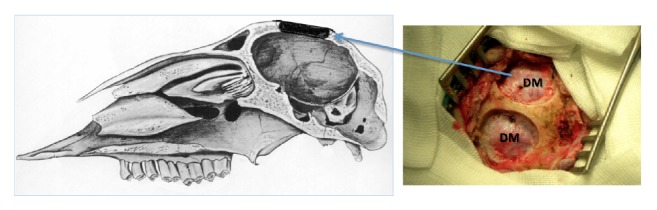
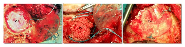
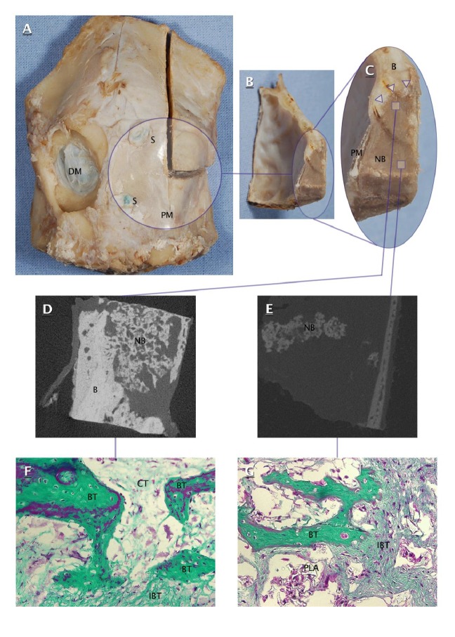
The aim of this study was to verify whether L-lactide/DL-lactide copolymer 80/20 (PLDLLA) and platelet-rich plasma (PRP) trigger bone formation within critical-sized calvarial defects in adult sheep (n = 6). Two craniectomies, each ca. 3 cm in diameter, were created in each animal. The first craniectomy was protected with an inner polylactide membrane, filled with PRP-polylactide granules, and covered with outer polylactide membrane. The second control craniectomy was left untreated. The animals were euthanized at 6, 7, 17, 19, 33, and 34 weeks after surgery, and the quality and the rate of reossification were assessed histomorphometrically and microtomographically. The study demonstrated that application of implants made of PLDLLA 80/20 combined with an osteopromotive substance (e.g., PRP) may promote bone healing in large calvarial defect in sheep. These promising proof-of-concept studies need to be verified in the future on a larger cohort of animals and over a longer period of time in order to draw definitive conclusions.


Figures

Figure 1
Figure 1
SEM microphotographs of polylactide membrane surface (a) and porous polylactide granule (b).
Figure 2
Figure 2
Sagittal cross section of ovine skull and intraoperative photograph of two calvarial craniectomies. DM: dura mater.
Figure 3
Figure 3
Consecutive stages of filling experimental craniectomy with polylactide implants and platelet-rich plasma (PRP). DM: dura mater; PG: polylactide granules mixed with PRP; PM: polylactide membrane.
Figure 4
Figure 4
Ovine calvarial specimen (A), transverse cross sections of filled craniectomy ((B) and (C)), microtomographic images of peripheral part (D) and central part (E) of filled craniectomy, and microphotographs of peripheral part (F) and central part (G) of filled craniectomy (200x, Masson's trichrome). DM: dura mater; S: suture; PM: polylactide membrane; B: calvarial bone; NB: newly formed bone in filled craniectomy; Δ: interface of newly formed bone and craniectomy edge; BT: bone trabecula; IBT: immature bone tissue; CT: connective tissue; PLA: polylactide.
Figure 5
Figure 5
Number of ossification foci (the sum of bone trabeculae and immature bone tissue) in the peripheral and central parts of the defect (Wilcoxon signed-rank test).
Figure 6
Figure 6
Area covered by newly formed bone (the sum of bone trabeculae and immature bone tissue) in the peripheral and central parts of the defect (Wilcoxon signed-rank test).
Figure 7
Figure 7
Area covered by connective tissue in the peripheral and central parts of the defect (Wilcoxon signed-rank test).
Figure 8
Figure 8
Correlation between follow-up time and total specimen area covered with newly formed bone (Spearman's rank correlation coefficient).

References

    1. Bobinski L., Koskinen L.-O. D., Lindvall P. Complications following cranioplasty using autologous bone or polymethylmethacrylate - Retrospective experience from a single center. 2013;115(9):1788–1791. doi: 10.1016/j.clineuro.2013.04.013. - DOI - PubMed
    1. Coulter I. C., Pesic-Smith J. D., Cato-Addison W. B., et al. Routine but risky: A multi-centre analysis of the outcomes of cranioplasty in the Northeast of England. 2014;156(7):1361–1368. doi: 10.1007/s00701-014-2081-1. - DOI - PubMed
    1. Piitulainen J. M., Kauko T., Aitasalo K. M. J., Vuorinen V., Vallittu P. K., Posti J. P. Outcomes of cranioplasty with synthetic materials and autologous bone grafts. 2015;83(5):708–714. doi: 10.1016/j.wneu.2015.01.014. - DOI - PubMed
    1. Bizenjima T., Takeuchi T., Seshima F., Saito A. Effect of poly (lactide-co-glycolide) (PLGA)-coated beta-tricalcium phosphate on the healing of rat calvarial bone defects: a comparative study with pure-phase beta-tricalcium phosphate. 2016;27(11):1360–1367. doi: 10.1111/clr.12744. - DOI - PubMed
    1. Melo L. G. N., Nagata M. J. H., Bosco A. F., Ribeiro L. L. G., Leite C. M. Bone healing in surgically created defects treated with either bioactive glass particles, a calcium sulfate barrier, or a combination of both materials: A histological and histometric study in rat tibias. 2005;16(6):683–691. doi: 10.1111/j.1600-0501.2005.01090.x. - DOI - PubMed

LinkOut - more resources