Review
. 2018 Aug;97(9):977-986.
doi: 10.1177/0022034518778033. Epub 2018 Jun 4.

A Review of the Impact of Implant Biomaterials on Osteocytes

Affiliations
Review

A Review of the Impact of Implant Biomaterials on Osteocytes

F A Shah et al. J Dent Res. 2018 Aug.

Abstract

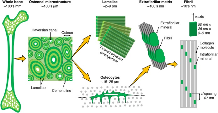
In lamellar bone, a network of highly oriented interconnected osteocytes is organized in concentric layers. Through their cellular processes contained within canaliculi, osteocytes are highly mechanosensitive and locally modulate bone remodeling. We review the recent developments demonstrating the significance of the osteocyte lacuno-canalicular network in bone maintenance around implant biomaterials. Drilling during implant site preparation triggers osteocyte apoptosis, the magnitude of which correlates with drilling speed and heat generation, resulting in extensive remodeling and delayed healing. In peri-implant bone, osteocytes physically communicate with implant surfaces via canaliculi and are responsive to mechanical loading, leading to changes in osteocyte numbers and morphology. Certain implant design features allow peri-implant osteocytes to retain a less aged phenotype, despite highly advanced extracellular matrix maturation. Physicochemical properties of anodically oxidized surfaces stimulate bone formation and remodeling by regulating the expression of RANKL (receptor activator of nuclear factor-κB ligand), RANK, and OPG (osteoprotegerin) from implant-adherent cells. Modulation of certain osteocyte-related molecular signaling mechanisms (e.g., sclerostin blockade) may enhance the biomechanical anchorage of implants. Evaluation of the peri-implant osteocyte lacuno-canalicular network should therefore be a necessary component in future investigations of osseointegration to more completely characterize the biological response to materials for load-bearing applications in dentistry and orthopedics.

Keywords: biocompatible materials; bone; bone matrix; bone-implant interface; dental implants; osseointegration.


Conflict of interest statement

The authors declare no potential conflicts of interest with respect to the authorship and/or publication of this article.

Figures

Figure 1.
Figure 1.
Bone consists of either a porous trabecular framework or a dense cortical structure. In cortical bone (e.g., in the middiaphysis of the femur), the microstructure consists of osteons (170- to 250-µm diameter), which are the units of bone produced during remodeling. Osteons contain a central vascular canal, the Haversian canal (60- to 90-μm diameter), concentrically surrounded by lamellae having a twisted plywood arrangement, where neighboring lamellae have different fibril orientations. Osteocytes reside in lacunae interconnected through canaliculi (100- to 400-nm diameter). Lamellae are composed of collagen fibrils (80- to 100-nm diameter). Fibrils are surrounded by extrafibrillar mineral platelets. Within the fibrils, type I collagen molecules and carbonated apatite crystallites form a nanocomposite structure.
Figure 2.
Figure 2.
The proximity of osteocytes (Ot) to implant surfaces can be observed with (a) histology and (b) backscattered electron scanning electron microscopy. Scale bars: 50 µm. CoCr, cobalt chromium. (c) Techniques such as resin cast etching allow visualization of osteocyte attachment to implant surfaces. Scale bar: 5 µm. Here, scanning electron microscopy images obtained in the secondary electron mode at 5 kV (red channel) and 20 kV (green channel) have been merged to achieve contrast. (Adapted with permission from Shah, Omar, et al. 2016. Copyright 2016, Elsevier.) (d) Osteocyte attachment to an acid-etched micrometer-smooth surface. Scale bar: 2 µm. (e) Interdigitation of canaliculi with the implant surface (arrowheads). Scale bar: 1 µm. (Reproduced from Shah, Stenlund, et al. 2016 under the terms of the Creative Commons Attribution License.) (f, g) Canaliculi (Ot.Ca) adherent to the surface of a laser-ablated implant. Scale bars: 200 nm (f), 1 µm (g). (h) Cross-sectional view of canaliculi (asterisks) in close association with the thickened-surface TiO2 layer of a laser-ablated implant (high-angle annular dark-field scanning transmission electron microscopy). Scale bar: 200 nm. (Reproduced from Shah, Johansson, et al. 2016 under the terms of the Creative Commons Attribution License.) (i) Osteocytes align parallel to the lamellar direction, which closely follows the microcontour of the laser-ablated implant surface. Scale bar: 100 µm. (j) Adjacent to the implant surface, osteocytes form an interconnected network. Scale bar: 25 µm. (k) Canaliculi (arrowheads) can be identified within the first several micrometers from the implant surface (high-angle annular dark-field scanning transmission electron microscopy). Scale bar: 1 µm. (l) Mineralized collagen fibrils appear to “wrap around” the canaliculus (asterisk). Scale bar: 200 nm. (Adapted with permission from Shah et al. 2014. Copyright 2014, Elsevier.)
Figure 3.
Figure 3.
Direct attachment of osteocytes to the implant surface. (a) Osteocytes (Ot) retain connectivity to the implant surface after 4 y of clinical function through canaliculi (arrowheads). (b) Ultrastructural similarities exist between the bone-implant interface and the bone-osteocyte interface (high-angle annular dark-field scanning transmission electron microscopy and electron tomography), where both comprise highly aligned collagen fibrils forming typical rope-like bundles. Scale bar: 1 µm. (Adapted with permission from Shah et al. 2015. Copyright 2015, American Chemical Society.) (c) Interconnected osteocyte lacuno-canalicular network within 60-µm-wide features on the surface of 3D printed Ti6Al4V. (d) An osteocyte (box in c) attaches to the implant surface through numerous branching canaliculi (one of which is indicated by an arrowhead). (e) Interconnected osteocytes within a 14-µm-wide crevice on the surface of 3D printed Ti6Al4V. (Adapted with permission from Shah, Snis, et al. 2016. Copyright 2016, Elsevier.) Scale bars: 10 µm (c, e), 5 µm (d). Pt, platinum; Ti, titanium.
Figure 4.
Figure 4.
Mechanical loading influences collagen alignment, osteocyte morphology, and the expression and production of osteocyte-related genes and proteins. (a, b) Polarized light microscopy reveals a preferential alignment of collagen fibers. The angle difference between groove direction and alignment direction of collagen fibers in +60° groove is smaller than in −60° groove. The grooves are 400 μm in pitch and 200 μm in depth. (Adapted with permission from Kuroshima et al. 2017. Copyright 2017, Elsevier.) (c–e) Assessment of osteocyte morphology via resin cast etching: (d) aspect ratio and (e) dendricity. (Adapted with permission from Sasaki et al. 2015. Copyright 2017, Elsevier.) Sclerostin immunoreactivity in peri-implant bone at (f) 5 d, (g) 10 d, (h) 20 d, (i) 2 mo, and (j) 7 mo. Scale bars: 35 µm. (Adapted with permission from Haga et al. 2011. Copyright 2011, John Wiley and Sons.) Influence of cyclic mechanical loading on the production of the osteoprotective gene Sema3A; as compared with (k) unloaded conditions, loading can increase (l) Sema3A production (arrowheads). Scale bars: 50 µm. (Reproduced from Uto et al. 2017 under the terms of the Creative Commons Attribution License.)
Figure 5.
Figure 5.
The impact of various factors—including implant physical and chemical properties (e.g., surface topography, bulk material), mechanical loading conditions, and healing time—on the morphology and resulting function of the osteocyte lacuno-canalicular system in the vicinity of implant biomaterials.

References

    1. Barros RR, Degidi M, Novaes AB, Piattelli A, Shibli JA, Iezzi G. 2009. Osteocyte density in the peri-implant bone of immediately loaded and submerged dental implants. J Periodontol. 80(3):499–504. - PubMed
    1. Buenzli PR, Sims NA. 2015. Quantifying the osteocyte network in the human skeleton. Bone. 75:144–150. - PubMed
    1. Cha JY, Pereira MD, Smith AA, Houschyar KS, Yin X, Mouraret S, Brunski JB, Helms JA. 2015. Multiscale analyses of the bone-implant interface. J Dent Res. 94(3):482–490. - PMC - PubMed
    1. Chen CH, Pei X, Tulu US, Aghvami M, Chen CT, Gaudilliere D, Arioka M, Maghazeh Moghim M, Bahat O, Kolinski M, et al. 2017. A comparative assessment of implant site viability in humans and rats. J Dent Res. 97(4):451–459. - PMC - PubMed
    1. Dallas SL, Prideaux M, Bonewald LF. 2013. The osteocyte: an endocrine cell . . . and more. Endocr Rev. 34(5):658–690. - PMC - PubMed

Publication types

LinkOut - more resources