

Mid-range probing-towards range-guided particle therapy
- PMID: 29864023
- PMCID: PMC6298607
- DOI: 10.1088/1361-6560/aaca1b
Mid-range probing-towards range-guided particle therapy
Abstract
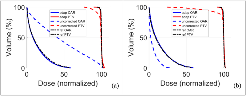
Particle therapy can achieve excellent dose localization but is sensitive to range uncertainty. Therefore, online in vivo range verification before treatment is critical for treatment safety and quality assurance. We introduce a novel range-probing technique that uses mid-range treatment spots selected from the treatment plan as probing beams to be delivered before other treatment spots in pencil beam scanning. The probing spot signal can be acquired by an in-beam positron emission tomography (PET) scanner, and the reconstructed spot positions are compared with pre-calculated positions to measure the range shift. Mid-range probing ensures that the Bragg peaks stay inside the tumor even with significant range variation from the plan. Single-layered spots enable easier spot detection than multi-layered spots without cross-layered spot smearing. With therapeutic dose, the probing beam offers higher positron activities and range detectability than the low-dose imaging beam by up to two orders of magnitude, without exposing patients to extra radiation. Higher positron activities allow sufficient signal statistics in shorter acquisition time, therefore reducing metabolic washout of positron emitters. Thus, range shifts from the plan can be measured easily. We also describe two online range-compensated plan modification methods. We apply correction, if the range shift is above a certain tolerance. We studied feasibility using simulated particle treatment plans with online anatomical changes. For illustration, we demonstrate range shift measurement using simulated probing dose. The proposed range probing and correction effectively handled range shifts in the simulated cases. Both range-compensated adaptation and optimization accounted for online changes so that the delivered dose matched the planned dose. With a dedicated online in-beam PET scanner and phantom and clinical studies, which are currently being developed, this novel strategy may open up a range-guided particle therapy1 paradigm.
Figures


References
-
- Assmann W, Kellnberger S, Reinhardt S, Lehrack S, Edlich A, Thirolf PG, Moser M, Dollinger G, Omar M, Ntziachristos V and Parodi K 2015. Ionoacoustic characterization of the proton Bragg peak with submillimeter accuracy Med Phys 42 (2) 567–574. - PubMed
-
- Attanasi F, Belcari N, Del Guerra A, Enghardt W, Moehrs S, Parodi K, Rosso V and Vecchio S 2009. Comparison of two dedicated ‘in beam’ PET systems via simultaneous imaging of C-12-induced beta(+)-activity Physics in Medicine and Biology 54 (2) N29–N35. - PubMed
-
- Dendooven P, Buitenhuis HJ, Diblen F, Heeres PN, Biegun AK, Fiedler F, van Goethem MJ, van der Graaf ER and Brandenburg S 2015. Short-lived positron emitters in beam-on PET imaging during proton therapy Phys Med Biol 60 (23) 8923–8947. - PubMed
-
- Dong L, Cheung JP and Zhu RX (2012) Image-Guided Proton and Carbon Ion Therapy Proton and Carbon Ion Therapy. Ma C and Lomax T, CRC Press: 127–149.
-
- Enghardt W, Crespo P, Fiedler F, Hinz R, Parodi K, Pawelke J and Ponisch F 2004. Charged hadron tumour therapy monitoring by means of PET Nuclear Instruments & Methods in Physics Research Section a-Accelerators Spectrometers Detectors and Associated Equipment 525 (1–2) 284–288.
Publication types
MeSH terms
Grants and funding
LinkOut - more resources
Full Text Sources
Other Literature Sources