. 2018 Jun 4;14(6):e1007059.
doi: 10.1371/journal.ppat.1007059. eCollection 2018 Jun.

Translational repression by an RNA-binding protein promotes differentiation to infective forms in Trypanosoma cruzi

Affiliations

Translational repression by an RNA-binding protein promotes differentiation to infective forms in Trypanosoma cruzi

Maria Albertina Romaniuk et al. PLoS Pathog. .

Abstract

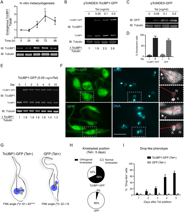
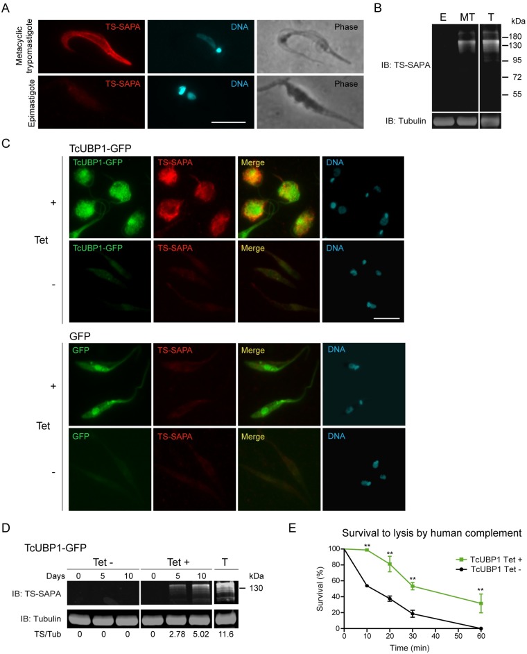
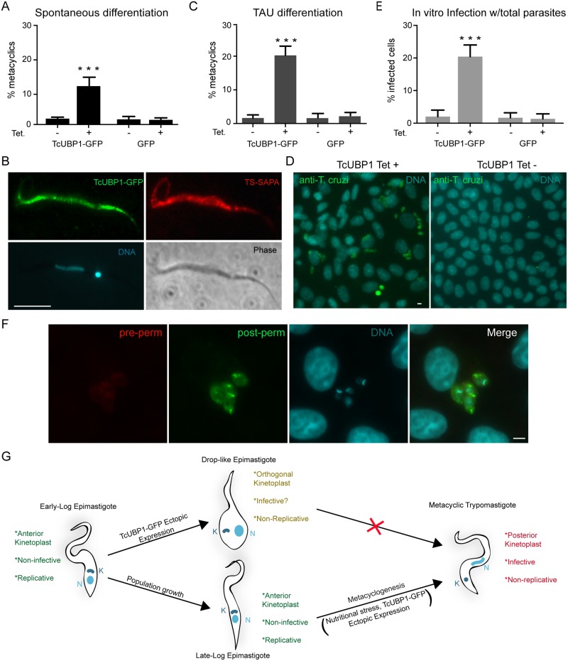
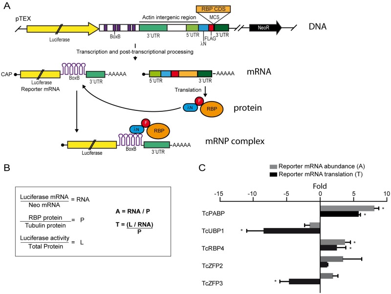
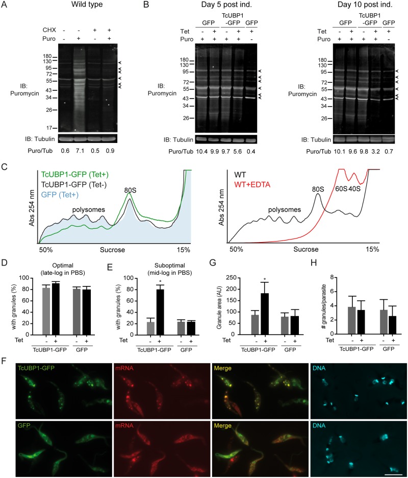
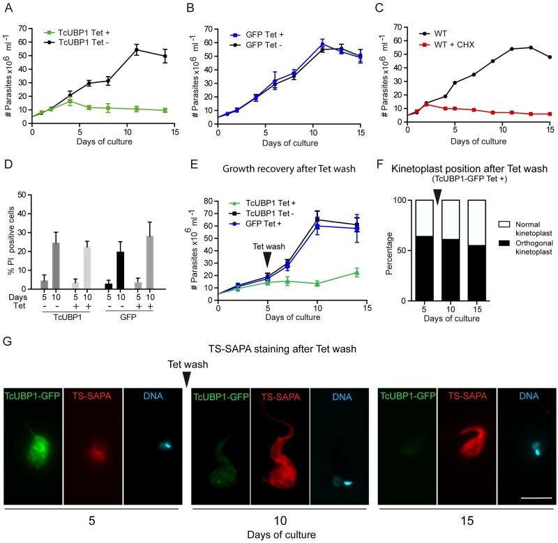
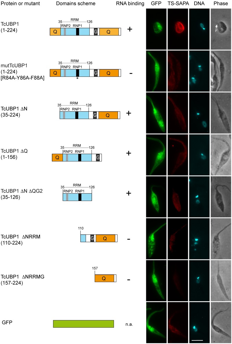
Trypanosomes, protozoan parasites of medical importance, essentially rely on post-transcriptional mechanisms to regulate gene expression in insect vectors and vertebrate hosts. RNA binding proteins (RBPs) that associate to the 3'-UTR of mature mRNAs are thought to orchestrate master developmental programs for these processes to happen. Yet, the molecular mechanisms by which differentiation occurs remain largely unexplored in these human pathogens. Here, we show that ectopic inducible expression of the RBP TcUBP1 promotes the beginning of the differentiation process from non-infective epimastigotes to infective metacyclic trypomastigotes in Trypanosoma cruzi. In early-log epimastigotes TcUBP1 promoted a drop-like phenotype, which is characterized by the presence of metacyclogenesis hallmarks, namely repositioning of the kinetoplast, the expression of an infective-stage virulence factor such as trans-sialidase, increased resistance to lysis by human complement and growth arrest. Furthermore, TcUBP1-ectopic expression in non-infective late-log epimastigotes promoted full development into metacyclic trypomastigotes. TcUBP1-derived metacyclic trypomastigotes were infective in cultured cells, and developed normally into amastigotes in the cytoplasm. By artificial in vivo tethering of TcUBP1 to the 3' untranslated region of a reporter mRNA we were able to determine that translation of the reporter was reduced by 8-fold, while its mRNA abundance was not significantly compromised. Inducible ectopic expression of TcUBP1 confirmed its role as a translational repressor, revealing significant reduction in the translation rate of multiple proteins, a reduction of polysomes, and promoting the formation of mRNA granules. Expression of TcUBP1 truncated forms revealed the requirement of both N and C-terminal glutamine-rich low complexity sequences for the development of the drop-like phenotype in early-log epimastigotes. We propose that a rise in TcUBP1 levels, in synchrony with nutritional deficiency, can promote the differentiation of T. cruzi epimastigotes into infective metacyclic trypomastigotes.


Conflict of interest statement

The authors have declared that no competing interests exist.

Figures

Fig 1
Fig 1. TcUBP1-GFP ectopic expression in epimastigotes.
(A) Wt epimastigotes were incubated in TAU-3AAG medium and TcUBP1 expression was assessed by Western blot for four days. Tubulin blot served as loading control. Values represent mean ± SD of 3 independent replicates. TcUBP1/Tubulin before TAU incubation was considered as 1. A representative Western blot experiment is shown. **P< 0.01 by Anova-Dunnett test. (B) Levels of TcUBP1-GFP induced expression 48 hours after the addition of different concentrations of Tet determined by Immunoblot (IB), and compared to TcUBP1 endogenous levels using an anti-TcUBP1 antibody. The ratio between the sum (Σ) of TcUBP1 forms (endogenous and ectopic) and Tubulin before Tet addition was considered as 1. (C) Levels of GFP induced expression at different Tet concentrations detected with an anti-GFP antibody and compared to Tubulin levels. (D) Percentage of GFP positive epimastigotes before and after induction with Tet determined by flow cytometry. Values represent mean ± SD of 6 independent replicates. (E) Immunoblot from a time course analysis of TcUBP1-GFP induced expression using 0.05 μg/ml Tet, together with endogenous TcUBP1. Tubulin was used as a loading control. The ratio between the sum of TcUBP1 forms (endogenous and ectopic) and Tubulin before Tet addition was considered as 1. (F) Fluorescence microscopy of epimastigotes expressing TcUBP1-GFP, or GFP, after 5 days of induction. Magnifications highlight a red line connecting the base of the flagellum, the nucleus and the kinetoplast, evidencing the repositioning of the kinetoplast. DNA was stained with DAPI, shown in cyan. Images are representative of 6 independent experiments. Scale bars, 5 μm. (G) Parasites schemes represent approximate forms obtained from induced TcUBP1-GFP or GFP cultures. In these parasites, the angle formed by the flagellum, the nucleus and the kinetoplast (FNK angle) was measured. The mean FNK angle is shown with black lines, and the kinetoplast is placed in this mean position. The gray spectrum represents SD above and below the mean FNK angle. ****P<0.0001 by T test (TcUBP1 N = 64, GFP N = 58). (H) Pie chart representing the mean percentages of cells with orthogonal or normal kinetoplast position in parasites induced for the expression of TcUBP1-GFP or GFP after 5 days. Values were obtained from 4 independent experiments. (I) Time course quantitation of parasites displaying drop-like phenotype in induced TcUBP1-GFP and GFP cultures.
Fig 2
Fig 2. Induced ectopic expression of TcUBP1-GFP in early-log phase epimastigotes promotes the development of metacyclic trypomastigotes features.
(A) Immunofluorescence of infective metacyclic trypomastigotes showing staining with anti-TS-SAPA antibody, and non-infective epimastigotes showing no staining. (B) Western blot of epimastigotes (E) and metacyclic trypomastigotes (MT) protein extracts with the anti-TS-SAPA antibody. An extract from cell-derived trypomastigotes (T) served as a positive control. Tubulin immunoblot was used as loading control. (C) Staining of TS-SAPA by immunofluorescence in induced (10 days) or uninduced early-log epimastigotes expressing TcUBP1-GFP or GFP. DNA was stained with DAPI. Images are representative of 3 independent experiments. (D) Western blot of uninduced or induced epimastigote protein extracts ectopically expressing TcUBP1-GFP. As a positive control for the anti-TS-SAPA antibody we used a protein extract from cell-derived trypomastigotes (T). Tubulin blot served as a loading control. Image is representative of 4 independent experiments. Scale bars, 5 μm. (E) Survival of epimastigotes to lysis by human complement. TcUBP1-GFP uninduced or induced parasites were incubated in PBS supplemented with 10% fresh human serum for the defined time course. Microscopic analysis was performed at each time point without fixation, only motile parasites were considered alive. **P< 0.01 by Two-way Anova- test.
Fig 3
Fig 3. TcUBP1-GFP ectopic expression in late-log phase epimastigotes promote full metacyclogenesis.
(A) The percentage of metacyclic trypomastigotes was determined in uninduced or induced TcUBP1-GFP or GFP late-log epimastigotes cultures 5 days after addition of Tet. Values represent mean ± SD of 4 independent replicates. (B) TS-SAPA staining was assessed in metacyclic trypomastigotes derived from TcUBP1-GFP induced cultures (5 days). Images are representative of 3 independent experiments. (C) The percentage of metacyclics was determined in uninduced or induced TcUBP1-GFP or GFP late-log epimastigotes incubated in TAU-3AAG medium for 96 hs. (D) Staining for intracellular amastigotes in VERO cells 7 days after incubating with induced and uninduced TcUBP1-GFP cultures. (E) Percentage of infected VERO cells 7 days post-infection with parasites derived from induced or uninduced TcUBP1-GFP or GFP cultures. Values represent mean ± SD of 3 independent replicates. (F) Infected VERO cells with intracellular amastigotes 7 days post infection with TcUBP1-GFP cultures. Amastigotes were stained with mouse anti-T. cruzi before permeabilization (red) and with rabbit anti-T. cruzi after permeabilization (green). DNA was stained with DAPI, shown in cyan. Images are representative of 3 independent experiments. ***P< 0.001 by Anova-Tukey test. Scale bars, 5 μm. (G) Model for TcUBP1-induced morphological differentiation.
Fig 4
Fig 4. Tethering of T. cruzi RBPs to reporter mRNA.
(A) Scheme of the DNA construct cloned in the pTEX vector. See Methods section for details. (B) Normalization of reporter mRNA and luciferase activity values. See Methods section for details. RNA: normalized luciferase mRNA, P: normalized RBP protein, L: normalized luciferase activity, A: reporter mRNA abundance, T: reporter mRNA translation. (C) Trypanosoma cruzi epimastigotes were transfected with the reporter construct in which different RBPs (TcPABP1, TcUBP1, TcRBP4, TcZFP2 and TcZFP3) were artificially tethered to the 3´ UTR of the reporter gene. The results indicate folds over values obtained with GFP as the tethered protein. Folds higher than 2 were considered significative (*). Results are shown for 3 or 4 independent experiments.
Fig 5
Fig 5. Translation repression in parasites ectopically expressing TcUBP1.
(A) Wt parasites were incubated with CHX (50 μg/ml) or vehicle and then with puromycin (10 μg/ml) or vehicle for 30 minutes. Puromycin incorporation was determined by Immunoblot (IB) using an anti-puromycin antibody. Protein loading was monitored using an anti-tubulin antibody. Image is representative of 3 independent experiments. Proteins unspecifically detected by the anti-puromycin antibody in the absence of puromycin, are pointed with arrowheads. (B) GFP or TcUBP1-GFP induced or GFP uninduced parasites were cultured for 5 and 10 days. 3 x 10⁷ parasites were incubated with puromycin (10 μg/ml) or vehicle for 30 minutes. Puromycin incorporation was determined by immunoblot using an anti-puromycin antibody. Protein loading was monitored using an anti-tubulin antibody. The ratio between Puromycin fluorescence/Tubulin fluorescence (Puro/Tub) is shown in arbitrary units (AU). Images are representative of 4 independent experiments. Unspecific bands are pointed with arrowheads. (C) Left panel, polysome profiles of induced (green line) and uninduced TcUBP1-GFP (black line), and from induced GFP parasites (filled light blue profile) after 5 days of induction, in the presence of CHX. Right panel, a control polysome profile from wt parasites using CHX is shown (black line), together with the disruption of 80S ribosomes into 60S and 40S ribosomal subunits with EDTA as a specificity control (red line). (D) Percentage of parasites displaying mRNA granules under optimal starvation conditions. (E) Percentage of parasites displaying mRNA granules under suboptimal starvation conditions. (F) Microscopic images of epimastigotes expressing TcUBP1-GFP, or GFP, under suboptimal starvation conditions, together with poly(A) mRNA detection by FISH and staining of DNA with DAPI, shown in cyan. Images are representative of 5 different experiments. (G) Analysis of granule area. (H) Analysis of the number of mRNA granules per parasite. Values represent mean ± SD of 5 independent replicates. *P< 0.05 by Anova-Dunnett test. Scale bar, 5 μm.
Fig 6
Fig 6. Irreversible growth arrest in parasites ectopically expressing TcUBP1.
(A and B) Growth curves of induced and non-induced epimastigotes expressing TcUBP1-GFP or GFP. Values represent mean ± SD of 5 independent replicates. (C) Growth curves of wt epimastigotes in the presence or absence of CHX. Values represent mean ± SD of 3 independent replicates. (D) The percentage of dead cells was determined by flow cytometry by incubating parasites with propidium iodide at 5 and 10 days after Tet addition. Values represent mean ± SD of 4 independent replicates. (E) Growth curves of induced and non-induced epimastigotes expressing TcUBP1-GFP or GFP. At day 5 of culture, Tet was removed by washing and fresh medium was added. Values represent mean ± SD of 4 independent replicates. (F) Kinetoplast repositioning was evaluated by microscopy. Cells were stained with DAPI. (G) Staining of TS-SAPA by immunofluorescence in induced early-log epimastigotes expressing TcUBP1-GFP, and after Tet wash. DNA was stained with DAPI, shown in cyan. Images are representative of 3 independent experiments.
Fig 7
Fig 7. Cellular morphology and TS-SAPA expression in parasites ectopically expressing mutated or truncated forms of TcUBP1.
Immunofluorescence of epimastigotes expressing the indicated TcUBP1-GFP constructs at day 10 of culture. Trans-sialidase expression was assessed using anti-TS-SAPA antibody and Alexa-568 goat anti-rabbit IgG. DNA was stained with DAPI shown in cyan. Images are representative of 3 independent experiments. Binding to RNA is based on previous evidence from our laboratory [19, 20]. Scale bars, 5 μm. Low complexity (LC) regions are shown in orange. RRM domain is shown in light blue.

Similar articles

Cited by

References

    1. Moore MJ, Proudfoot NJ. Pre-mRNA processing reaches back to transcription and ahead to translation. Cell. 2009;136(4):688–700. doi: 10.1016/j.cell.2009.02.001 . - DOI - PubMed
    1. Clery A, Allain FH. From Structure to Function of RNA Binding Domains In: RNA Binding Proteins. 2012;Chapter 9:137–58.
    1. Calabretta S, Richard S. Emerging Roles of Disordered Sequences in RNA-Binding Proteins. Trends in biochemical sciences. 2015;40(11):662–72. doi: 10.1016/j.tibs.2015.08.012 . - DOI - PubMed
    1. Kolev NG, Ullu E, Tschudi C. The emerging role of RNA-binding proteins in the life cycle of Trypanosoma brucei. Cellular microbiology. 2014;16(4):482–9. doi: 10.1111/cmi.12268 - DOI - PMC - PubMed
    1. Clayton C, Shapira M. Post-transcriptional regulation of gene expression in trypanosomes and leishmanias. Molecular and biochemical parasitology. 2007;156(2):93–101. doi: 10.1016/j.molbiopara.2007.07.007 . - DOI - PubMed

Publication types

MeSH terms