

Impact of euthanasia, dissection and postmortem delay on metabolic profile in mouse retina and RPE/choroid
- PMID: 29864440
- PMCID: PMC6110973
- DOI: 10.1016/j.exer.2018.05.032
Impact of euthanasia, dissection and postmortem delay on metabolic profile in mouse retina and RPE/choroid
Abstract
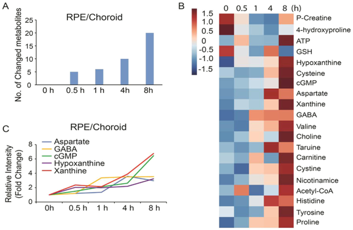
Metabolomics studies in the retina and retinal pigment epithelium (RPE) in animal models or postmortem donors are essential to understanding the retinal metabolism and to revealing the underlying mechanisms of retinal degenerative diseases. We have studied how different methods of euthanasia (CO2 or cervical dislocation) different isolation procedures and postmortem delay affect metabolites in mouse retina and RPE/choroid using LC MS/MS and GC MS. Compared with cervical dislocation, CO2 exposure for 5 min dramatically degrades ATP and GTP into purine metabolites in the retina while raising intermediates in glucose metabolism and amino acids in the RPE/choroid. Isolation in cold buffer containing glucose has the least change in metabolites. Postmortem delay time-dependently and differentially impacts metabolites in the retina and RPE/choroid. In the postmortem retina, 18% of metabolites were changed at 0.5 h (h), 41% at 4 h and 51% at 8 h. However, only 6% of metabolites were changed in the postmortem RPE/choroid and it steadily increased to 20% at 8 h. Notably, both postmortem retina and RPE/choroid tissue showed increased purine metabolites. Storage of eyes in cold nutrient-rich medium substantially blocked the postmortem change in the retina and RPE/choroid. In conclusion, our study provides optimized methods to prepare fresh or postmortem retina and RPE/choroid tissue for metabolomics studies.
Keywords: Euthanasia; Metabolite; Postmortem; RPE; Retina.
Copyright © 2018. Published by Elsevier Ltd.
Conflict of interest statement
Conflicts of interest
None declared.
Figures


Similar articles
-
Dissection of Human Retina and RPE-Choroid for Proteomic Analysis.J Vis Exp. 2017 Nov 12;(129):56203. doi: 10.3791/56203. J Vis Exp. 2017. PMID: 29155757 Free PMC article.
-
The retina and retinal pigment epithelium differ in nitrogen metabolism and are metabolically connected.J Biol Chem. 2020 Feb 21;295(8):2324-2335. doi: 10.1074/jbc.RA119.011727. Epub 2020 Jan 17. J Biol Chem. 2020. PMID: 31953322 Free PMC article.
-
Effect of acute and chronic aldosterone exposure on the retinal pigment epithelium-choroid complex in rodents.Exp Eye Res. 2019 Oct;187:107747. doi: 10.1016/j.exer.2019.107747. Epub 2019 Aug 5. Exp Eye Res. 2019. PMID: 31394103
-
Retina Metabolism and Metabolism in the Pigmented Epithelium: A Busy Intersection.Annu Rev Vis Sci. 2021 Sep 15;7:665-692. doi: 10.1146/annurev-vision-100419-115156. Epub 2021 Jun 8. Annu Rev Vis Sci. 2021. PMID: 34102066 Free PMC article. Review.
-
Retinal pigment epithelium lipid metabolic demands and therapeutic restoration.Taiwan J Ophthalmol. 2021 Aug 28;11(3):216-220. doi: 10.4103/tjo.tjo_31_21. eCollection 2021 Jul-Sep. Taiwan J Ophthalmol. 2021. PMID: 34703736 Free PMC article. Review.
Cited by
-
Metabolomics and biomarkers in ocular matrix: beyond ocular diseases.Int J Ophthalmol. 2020 Jun 18;13(6):991-1003. doi: 10.18240/ijo.2020.06.21. eCollection 2020. Int J Ophthalmol. 2020. PMID: 32566514 Free PMC article. Review.
-
Revival of light signalling in the postmortem mouse and human retina.Nature. 2022 Jun;606(7913):351-357. doi: 10.1038/s41586-022-04709-x. Epub 2022 May 11. Nature. 2022. PMID: 35545677 Free PMC article.
-
The Symbiotic Relationship between the Neural Retina and Retinal Pigment Epithelium Is Supported by Utilizing Differential Metabolic Pathways.iScience. 2020 Apr 24;23(4):101004. doi: 10.1016/j.isci.2020.101004. Epub 2020 Mar 21. iScience. 2020. PMID: 32252018 Free PMC article.
-
Reduced nuclear NAD+ drives DNA damage and subsequent immune activation in the retina.Hum Mol Genet. 2022 May 4;31(9):1370-1388. doi: 10.1093/hmg/ddab324. Hum Mol Genet. 2022. PMID: 34750622 Free PMC article.
-
Improved immunohistochemical detection of phosphorylated mitogen-activated protein kinases in the injured rat optic nerve head.Histochem Cell Biol. 2019 May;151(5):435-456. doi: 10.1007/s00418-019-01771-x. Epub 2019 Mar 11. Histochem Cell Biol. 2019. PMID: 30859291
References
-
- Ait-Ali N, Fridlich R, Millet-Puel G, Clerin E, Delalande F, Jaillard C, Blond F, Perrocheau L, Reichman S, Byrne LC, Olivier-Bandini A, Bellalou J, Moyse E, Bouillaud F, Nicol X, Dalkara D, van Dorsselaer A, Sahel JA, Leveillard T, 2015. Rod-derived cone viability factor promotes cone survival by stimulating aerobic glycolysis. Cell 161, 817–832. - PubMed
-
- Anderson RE, Maude MB, McClellan M, Matthes MT, Yasumura D, LaVail MM, 2002. Low docosahexaenoic acid levels in rod outer segments of rats with P23H and S334ter rhodopsin mutations. Mol. Vis 8, 351–358. - PubMed
-
- Belanger MP, Askin N, Wittnich C, 2002. Multiple in vivo liver biopsies using a freeze-clamping technique. J. Invest. Surg.: Offc. J. Acad. Surg. Res 15, 109–112. - PubMed
-
- Benveniste H, Drejer J, Schousboe A, Diemer NH, 1984. Elevation of the extra-cellular concentrations of glutamate and aspartate in rat hippocampus during transient cerebral ischemia monitored by intracerebral microdialysis. J. Neurochem 43, 1369–1374. - PubMed
-
- Brooks SP, Lampi BJ, Bihun CG, 1999. The influence of euthanasia methods on rat liver metabolism. Contemp. Top. Lab. Anim. Sci 38, 19–24. - PubMed
Publication types
MeSH terms
Substances
Grants and funding
LinkOut - more resources
Full Text Sources
Other Literature Sources
Miscellaneous
