. 2018 Jun 5;49(1):46.
doi: 10.1186/s13567-018-0540-z.

Elucidation of the pathology and tissue distribution of Lagovirus europaeus GI.2/RHDV2 (rabbit haemorrhagic disease virus 2) in young and adult rabbits (Oryctolagus cuniculus)

Affiliations

Elucidation of the pathology and tissue distribution of Lagovirus europaeus GI.2/RHDV2 (rabbit haemorrhagic disease virus 2) in young and adult rabbits (Oryctolagus cuniculus)

Aleksija Neimanis et al. Vet Res. .

Abstract

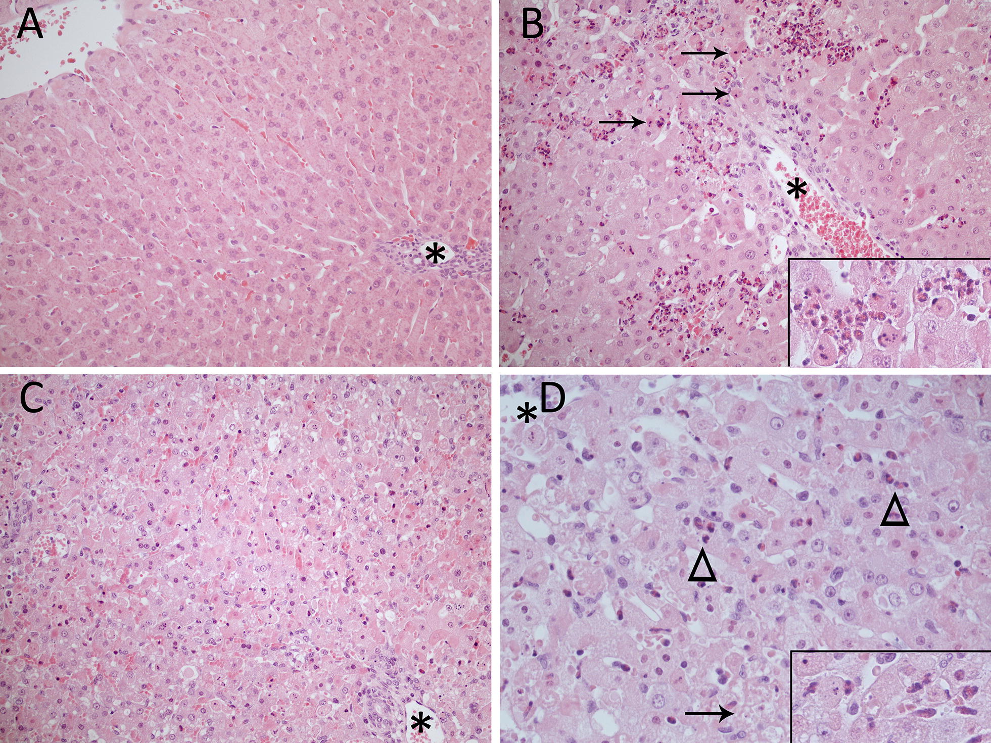
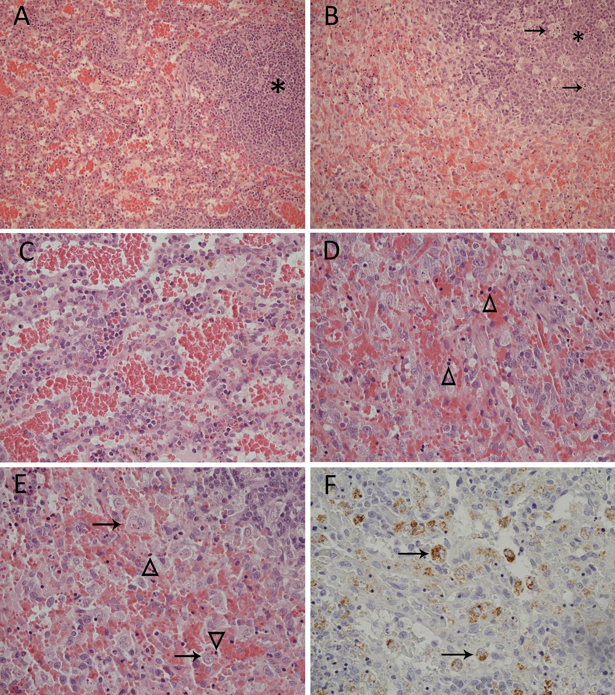
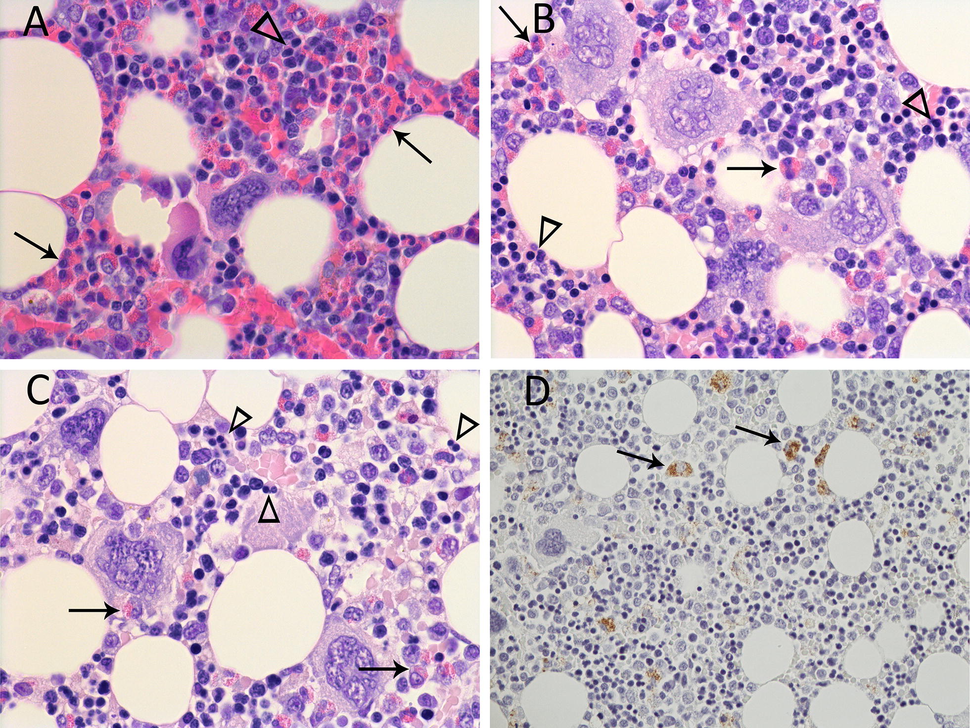
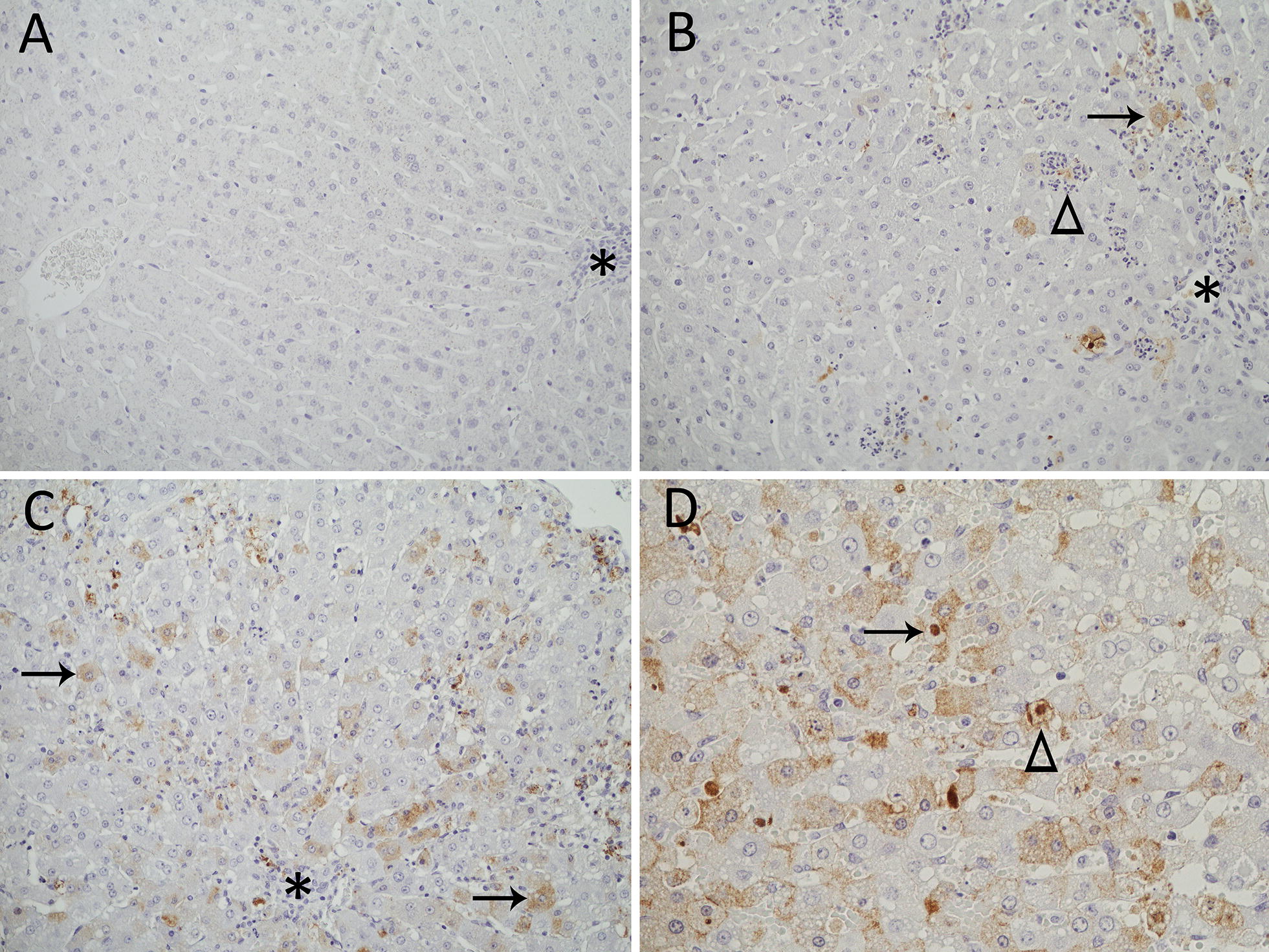
Lagovirus europaeus GI.2, also known as RHDV2 or RHDVb, is an emerging virus that causes rabbit haemorrhagic disease (RHD) in European rabbits (Oryctolagus cuniculus). In contrast to L. europaeus GI.1 (or RHDV/RHDVa) viruses that are only pathogenic for adults, GI.2 causes clinical disease in both adults and kittens. However, detailed descriptions of the pathology of this virus that may provide insight into its pathogenicity and emergence are lacking. Using an Australian GI.2 field strain isolated in 2015, we provide the first detailed description of pathology, viral antigen distribution and tissue load of GI.2 in adult and 5-week old New Zealand white rabbits using histology, immunohistochemistry and RT-qPCR. Liver was the target organ, but in contrast to GI.1 viruses, lesions and inflammatory responses did not differ between adults and kittens. Lymphocytic inflammation, proposed to be protective in kittens infected with GI.1, was notably absent. We also present the first descriptions of bone marrow changes in RHD, including decreased myeloid-to-erythroid ratio. Consistent with other pathogenic lagoviruses, intracellular viral antigen was demonstrated in hepatocytes and cells of the mononuclear phagocytic system. In terminal stages of disease, viral loads were highest in liver, serum and spleen. Despite the small sample size, our data suggest that unlike early European GI.2 strains, the pathogenicity of the Australian GI.2 virus is similar to GI.1 viruses. Additionally, GI.2 was fatal for all (n = 5) inoculated kittens in this study. This may significantly alter RHD epidemiology in the field, and may impact biocontrol programs for invasive rabbits in Australia where GI.1 viruses are intentionally released.


Figures

Figure 1
Figure 1
Microscopic lesions in the liver of rabbits (Oryctolagus cuniculus) inoculated with Lagovirus europaeus G1.2 (BlMt-1). Asterisks denote portal areas. Haematoxylin and eosin stain. A Uninfected control rabbit (rabbit 130). 200× magnification. B Earlier stage of acute infection in an adult rabbit (rabbit 283). There is degeneration and death of hepatocytes at the limiting plate and periportally surrounded by intensive heterophilic inflammation. Arrows denote apoptotic cells. 200× magnification. Insert: high magnification of dead hepatocytes surrounded by heterophils. C Terminal stage of acute infection in a 5 week old rabbit (rabbit 303). There is massive hepatocellular degeneration and death. 200× magnification. D Terminal stage of acute infection in a 5 week old rabbit (rabbit 306). Arrowheads denote clusters of heterophils that are seen throughout the lobule and the arrow indicates an area of lytic necrosis. 400× magnification. Insert: high magnification of heterophils surrounding dead hepatocytes.
Figure 2
Figure 2
Microscopic lesions and viral antigen localization in the spleen of a 5 week old rabbit (Oryctolagus cuniculus) (rabbit 306) inoculated with Lagovirus europaeus G1.2 (BlMt-1). Asterisks denote the white pulp. A Uninfected control (rabbit 130). 200× magnification. B Terminal stage of acute infection. There is lymphoid depletion and tingible body macrophages (arrows) in the white pulp and macrophage hyperplasia in the adjacent red pulp. 200× magnification. C Red pulp of the uninfected control (rabbit 130). 400× magnification. D Red pulp of rabbit 306. Note the indistinct, hyaline stroma and presence of small, dark pyknotic cells (arrowheads). 400× magnification. E Rabbit 306. Active phagocytosis by macrophages in the red pulp (arrows) and numerous pyknotic cells (arrowheads). The macrophage indicated at the bottom of the image has phagocytized a pyknotic cell. 400× magnification. F Immunohistochemical visualization of viral capsid antigen (brown staining) in the cytoplasm of viable macrophages (arrows) in the terminal stage of acute infection. Note that no staining is associated with pyknotic cells. 400× magnification.
Figure 3
Figure 3
Microscopic lesions and viral antigen localization in the bone marrow of adult rabbits (Oryctolagus cuniculus) inoculated with Lagovirus europaeus G1.2 (BlMt-1). Arrows denote myeloid cells and arrowheads denote erythroid cells. A Uninfected control (rabbit 130). 600× magnification. B Bone marrow in an earlier stage of acute infection (rabbit 283). Note the decreasing proportion of myeloid to erythroid cells. 600× magnification. C Bone marrow at the terminal stage of acute infection (rabbit 282). Myeloid cells are scarce and erythroid cells at arrowheads are binucleated. 600× magnification. D Immunohistochemical visualization of viral capsid antigen (brown staining) in the cytoplasm of viable macrophages (arrows) in the terminal stage of acute infection (rabbit 282). 400× magnification.
Figure 4
Figure 4
Viral antigen localization in the liver of rabbits (Oryctolagus cuniculus) inoculated with Lagovirus europaeus G1.2 (BlMt-1). Asterisks denote portal areas. A Uninfected control (rabbit 130). No antigen (brown staining) is seen. 200× magnification. B Earlier stage of acute infection in an adult rabbit (rabbit 283). A small number of periportal hepatocytes contain viral antigen (brown staining, arrow). A cluster of heterophils surrounds necrotic, antigen-containing debris interpreted to be the remnants of an infected hepatocyte (arrowhead). 200× magnification. C Terminal stage of acute infection in an adult rabbit (rabbit 282). There is cytoplasmic staining in hepatocytes throughout the lobule (arrows). 200× magnification. D Terminal stage of acute infection in a 5 week old rabbit (rabbit 303). In addition to the finely stippled intracytoplasmic staining of infected hepatocytes, there is occasional intranuclear staining (arrow, arrowhead) and staining of the cell membrane (arrowhead). 400× magnification.

References

    1. Abrantes J, van der Loo W, Le Pendu J, Esteves PJ. Rabbit haemorrhagic disease (RHD) and rabbit haemorrhagic disease virus (RHDV): a review. Vet Res. 2012;43:12. doi: 10.1186/1297-9716-43-12. - DOI - PMC - PubMed
    1. Le Gall-Reculé G, Lavazza A, Marchandeau S, Bertagnoli S, Zwingelstein F, Cavadini P, Martinelli N, Lombardi G, Guerin J-L, Lemaitre E, Decors A, Boucher S, Le Normand B, Capucci L. Emergence of a new lagovirus related to rabbit haemorrhagic disease virus. Vet Res. 2013;44:81. doi: 10.1186/1297-9716-44-81. - DOI - PMC - PubMed
    1. Le Pendu J, Abrantes J, Bertagnoli S, Guitton JS, Le Gall-Reculé G, Lopes AM, Marchandeau S, Alda F, Almeida T, Célio AP, Bárcena J, Burmakina G, Blanco E, Calvete C, Cavadini P, Cooke B, Dalton K, Delibes Mateos M, Deptula W, Eden JS, Wang F, Ferreira CC, Ferreira P, Foronda P, Gonçalves D, Gavier-Widén D, Hall R, Hukowska-Szematowicz B, Kerr P, Kovaliski J, et al. Proposal for a unified classification system and nomenclature of lagoviruses. J Gen Virol. 2017;98:1658–1666. doi: 10.1099/jgv.0.000840. - DOI - PubMed
    1. Le Gall-Reculé G, Zwingelstein F, Boucher S, Le Normand B, Plassiart G, Portejoie Y, Decors A, Bertagnoli S, Guerin JL, Marchandeau S. Detection of a new variant of rabbit haemorrhagic disease virus in France. Vet Rec. 2011;168:137–138. doi: 10.1136/vr.d697. - DOI - PubMed
    1. Dalton KP, Nicieza I, Balseiro A, Muguerza MA, Rosell JM, Casais R, Alvarez AL, Parra F. Variant rabbit hemorrhagic disease virus in young rabbits, Spain. Emerg Infect Dis. 2012;18:2009–2012. doi: 10.3201/eid1812.120341. - DOI - PMC - PubMed

Publication types

LinkOut - more resources