Review
. 2018 Jul 12;132(2):159-169.
doi: 10.1182/blood-2018-02-769026. Epub 2018 Jun 4.

Activated protein C, protease activated receptor 1, and neuroprotection

Affiliations
Review

Activated protein C, protease activated receptor 1, and neuroprotection

John H Griffin et al. Blood. .

Abstract

Protein C is a plasma serine protease zymogen whose active form, activated protein C (APC), exerts potent anticoagulant activity. In addition to its antithrombotic role as a plasma protease, pharmacologic APC is a pleiotropic protease that activates diverse homeostatic cell signaling pathways via multiple receptors on many cells. Engineering of APC by site-directed mutagenesis provided a signaling selective APC mutant with 3 Lys residues replaced by 3 Ala residues, 3K3A-APC, that lacks >90% anticoagulant activity but retains normal cell signaling activities. This 3K3A-APC mutant exerts multiple potent neuroprotective activities, which require the G-protein-coupled receptor, protease activated receptor 1. Potent neuroprotection in murine ischemic stroke models is linked to 3K3A-APC-induced signaling that arises due to APC's cleavage in protease activated receptor 1 at a noncanonical Arg46 site. This cleavage causes biased signaling that provides a major explanation for APC's in vivo mechanism of action for neuroprotective activities. 3K3A-APC appeared to be safe in ischemic stroke patients and reduced bleeding in the brain after tissue plasminogen activator therapy in a recent phase 2 clinical trial. Hence, it merits further clinical testing for its efficacy in ischemic stroke patients. Recent studies using human fetal neural stem and progenitor cells show that 3K3A-APC promotes neurogenesis in vitro as well as in vivo in the murine middle cerebral artery occlusion stroke model. These recent advances should encourage translational research centered on signaling selective APC's for both single-agent therapies and multiagent combination therapies for ischemic stroke and other neuropathologies.


Conflict of interest statement

Conflict-of-interest disclosure: B.V.Z. is a founder of ZZ Biotech LLC, a biotechnology company with a mission to develop APC and its functional mutants for the treatment of stroke and other neurological disorders. L.O.M. and J.H.G. are inventors for some uses of APC mutants, and J.H.G. is a consultant for ZZ Biotech LLC.

Figures
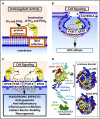
Figure 1.
Figure 1.
APC anticoagulant and cell signaling pathways and the structure of signaling-selective 3K3A-APC. (A) Anticoagulant activity of APC involves the proteolytic inactivation of factors Va and VIIIa on membrane surfaces containing phospholipids that are derived from cells, platelets, lipoproteins, or cellular microparticles. The irreversible inactivation of factors Va and VIIIa to yield inactive factors Vi and VIIIi by APC is accelerated by a variety of lipid and protein cofactors (eg, glucosyl ceramide, protein S, etc). (B) Beneficial direct effects of APC on cells require the EPCR and PAR1. One distinction between proinflammatory thrombin signaling and cytoprotective APC signaling is the localization of APC signaling in the caveolin-1–rich microdomains (caveolae). (C) Neuroprotective mechanisms for APC effects on cells may also involve other receptors including PAR-3. APC-initiated signaling effects on cells can include antiapoptotic activities, anti-inflammatory activities, inhibition of the inflammasome, stabilization of endothelial barrier functions including the BBB, and neurogenesis. (D) The polypeptide structure of APC comprises an N-terminal GLA domain (green) that binds to negatively charged lipids and EPCR, 2 EGF-like domains (blue), and the protease domain containing the active site triad of serine, histidine, and aspartic acid residues (red). Four glycosylation sites are indicated by gray-shaded moieties. Substrate selectivity of this protease is determined by interactions between the targeted substrates and the active site and also by multiple unique binding exosites on APC that vary for different substrates. The protease domain space–filled model (see insert) highlights in the yellow box 3 positively charged lysine (K) residues within the so-called 37 loop (KKK 191-193), which is an exosite for APC’s recognition of factors Va and VIIIa. Mutation of these 3 residues to alanine (3K3A-APC) reduces APC’s anticoagulant activity by >90%, but it does not affect its interactions with the cytoprotective substrates, PAR1, or its other known cell signaling receptors. Thus, 3K3A-APC is very signaling selective.
Figure 2.
Figure 2.
APC causes biased signaling via PAR1 cleavage at Arg46. PAR1 is a 7-transmembrane GPCR receptor capable of many conformational states. (A) The extracellular N-terminus contains the intramolecular ligands, which become exposed after proteolysis by APC or thrombin. PAR1 cleavage by thrombin at Arg41 exposes the canonical N-terminal tethered agonist that begins with residue Ser42 (SFLLRN-), whereas noncanonical cleavage by APC at Arg46 results in a different N-terminal tethered agonist that begins with residue Asn47 (NPNDKY-). (B) Synthetic agonist peptides with the N-terminal tethered-ligand sequences beginning with residue 42 (TRAP) or residue 47 (TR47) cause thrombin-like or APC-like effects on cells, respectively. (C) Activation of PAR1 by thrombin or TRAP stabilizes PAR1 conformers, whose intracellular loops provide surfaces that are highly favorable for interactions with G proteins, resulting in G-protein–dependent signaling. These PAR1 conformers are termed “G-protein biased.” In contrast, activation of PAR1 by APC or TR47 stabilizes different PAR1 conformers that preferentially interact with β-arrestin-2, resulting in β-arrestin-2–dependent signaling. Such PAR1 conformers are termed “β-arrestin biased.” Thus, the agonist bias is directly related to the cleavage site used to activate PAR1 because the cleavage determines which tethered ligand is exposed and which subsets of PAR1 conformations are stabilized. Biased agonism results in the induction of uniquely different signaling repertoires, such as the activation of ERK1/2 and RhoA, etc, resulting in vascular leakage by thrombin or the activation of PI3K, Akt, and Rac1, etc, by APC resulting in neuroprotection. (D-E) APC-mediated neuroprotection in ischemic stroke requires PAR1-dependent biased signaling due to cleavage at Arg46 in PAR1. To assess PAR1-biased signaling, studies employed homozygous QQ41-PAR1 mice, homozygous QQ46-PAR1 mice, and wt control mice. At 4 hours after a 60-minute MCAO, 3K3A-APC (0.04 mg/kg) or placebo was given IV, and then at 24 hours after occlusion, various parameters were measured. Treatment of mice with 3K3A-APC or vehicle is indicated by plus or negative signs under panels D and E. Data are shown for infarct volume (D) and for degenerating neurons as determined by Fluoro-Jade C stain (E) for each mouse group. For panels D and E, bars indicate mean ± standard deviation, n = 4-6 mice per group. (For details regarding panels D and E, see Sinha et al).
Figure 3.
Figure 3.
Neuroprotective effects of APC in the neurovascular space and in neurons. (A) APC can provide multiple neuroprotective effects in the neurovascular unit of the brain after ischemic stroke. APC inhibits the breakdown of the BBB, preventing extravasation of inflammatory cells. EPCR-mediated transfer of APC across the BBB permits APC to engage PAR1, PAR3, and EPCR directly on neurons, glia, and other cells in the brain to convey multiple cytoprotective activities and dampen neuronal damage. APC attenuates neuroinflammatory responses. Furthermore, APC promotes neurogenesis and vascular regeneration in the brain that directly contribute to repair and regeneration of the affected brain tissue after ischemic stroke (see Figure 4). Studies showing the requirement for PAR1 for APC’s neuroprotection are seen in panels B-D that show an assessment of brain damage after a 1-hour transient MCAO in wt (PAR1+/+) and knockout (PAR1−/−) mice treated with recombinant murine (rm)-tPA and recombinant murine wt-APC (0.2 mg/kg). Damage quantified at 24 hours after onset of ischemia was based on brain infarct volume (B), hemorrhage (C), and altered levels of NF-ĸB (D). Values are mean ± standard error of the mean (SEM), and n = 3-6 mice per group; * designates data for mice receiving both APC and rm-tPA (for details regarding panels B-D, see Cheng et al). (E) Studies using cultured neuronal cells from wt mice (PAR1+/+, PAR3+/+) and PAR1−/− and PAR3−/− knockout mice treated with 3K3A-APC showed the requirement for both PAR1 and PAR3 for 3K3A-APC’s direct neuronal protection against N-methyl-d-aspartate–induced excitotoxic injury of neurons (E). Values are mean ± SEM, n = 5 mice per group. (For details regarding panel E, see Guo et al).
Figure 4.
Figure 4.
Regenerative activities of signaling-selective 3K3A-APC for human NSCs. (A-C) 3K3A-APC stimulates neuronal proliferation and differentiation from human embryo-derived NSCs in vitro. (A) Stimulation of human NSCs proliferation in culture by 3K3A-APC requires PAR1 and PAR3 but not PAR2. Quantification of proliferation was based on the percentage of Ki-67+/nestin+ cells in culture after 48 hours in the presence and absence of 3K3A-APC (2 nM) and/or PAR1-, PAR2-, and PAR3-specific cleavage site blocking antibodies. (B-C) Enhancement of neuronal proliferation and differentiation of NSCs by 3K3A-APC requires activation of Akt. Activation of the PI3K/Akt pathway by human recombinant 3K3A-APC (2 nM) in human NSCs requires PAR1 and PAR3 (B) and SphK-1 but not SphK-2 (C). Phosphorylation of Akt at Ser473 (pAkt) and total Akt was determined 3 hours after 3K3A-APC or vehicle treatment by western blot in whole-cell extracts of NSCs transfected with PAR1, PAR3, SphK-1, SphK-2, or control small interfering RNA. Intensity of pAkt signal was determined by scanning densitometry and normalized to total Akt. Data are shown as mean ± SEM, n = 3 independent cultures in triplicate. Statistical significance was determined by 1-way analysis of variance followed by Tukey’s post hoc test. NS, not significant. (For details regarding panels A-C, see Guo et al). (D) Schematic overview of APC’s regenerative activities for human NPCs. Activation of the PI3K/Akt pathway in NSCs by 3K3A-APC requires PAR1, PAR3, S1P1, SphK-1, and EPCR. Through integration of signaling linked to multiple downstream effectors, activation of the PI3K/Akt signaling node by 3K3A-APC induces proliferation, migration, survival, and differentiation of NSCs. SphK-1, sphingosine kinase-1; SphK-2, sphingosine kinase-2.

References

    1. Seligsohn U, Lubetsky A. Genetic susceptibility to venous thrombosis. N Engl J Med. 2001;344(16):1222-1231. - PubMed
    1. Branson HE, Katz J, Marble R, Griffin JH. Inherited protein C deficiency and coumarin-responsive chronic relapsing purpura fulminans in a newborn infant. Lancet. 1983;2(8360):1165-1168. - PubMed
    1. Goldenberg NA, Manco-Johnson MJ. Protein C deficiency. Haemophilia. 2008;14(6):1214-1221. - PubMed
    1. Chalmers E, Cooper P, Forman K, et al. . Purpura fulminans: recognition, diagnosis and management. Arch Dis Child. 2011;96(11):1066-1071. - PubMed
    1. Ohga S, Ishiguro A, Takahashi Y, et al. ; Japan Childhood Thrombophilia Study Group. Protein C deficiency as the major cause of thrombophilias in childhood. Pediatr Int. 2013;55(3):267-271. - PubMed

Publication types

MeSH terms