. 2018 Jun 19;115(25):6488-6493.
doi: 10.1073/pnas.1721769115. Epub 2018 Jun 4.

Uncovering pH at both sides of the root plasma membrane interface using noninvasive imaging

Affiliations

Uncovering pH at both sides of the root plasma membrane interface using noninvasive imaging

Alexandre Martinière et al. Proc Natl Acad Sci U S A. .

Abstract

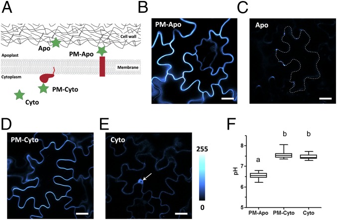
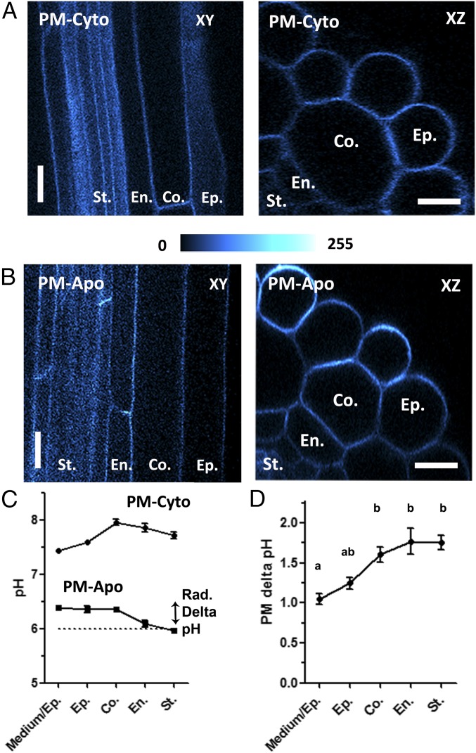
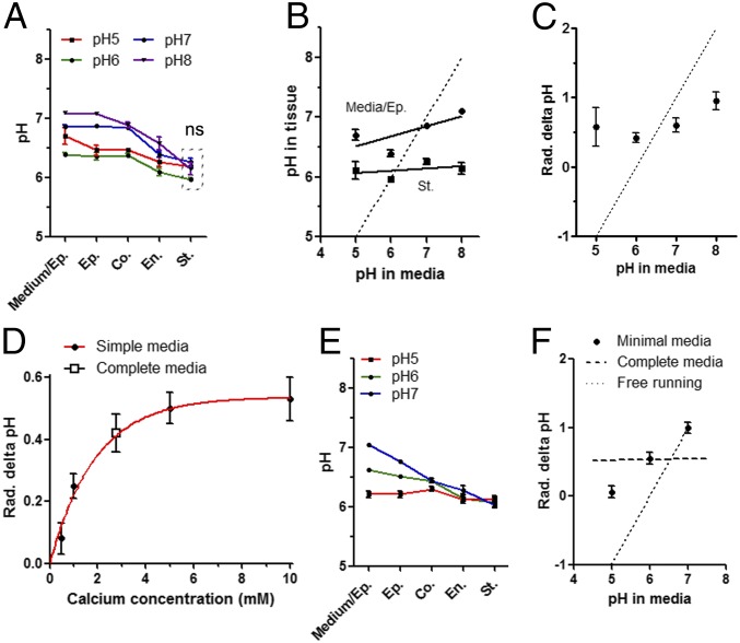
Building a proton gradient across a biological membrane and between different tissues is a matter of great importance for plant development and nutrition. To gain a better understanding of proton distribution in the plant root apoplast as well as across the plasma membrane, we generated Arabidopsis plants expressing stable membrane-anchored ratiometric fluorescent sensors based on pHluorin. These sensors enabled noninvasive pH-specific measurements in mature root cells from the medium-epidermis interface up to the inner cell layers that lie beyond the Casparian strip. The membrane-associated apoplastic pH was much more alkaline than the overall apoplastic space pH. Proton concentration associated with the plasma membrane was very stable, even when the growth medium pH was altered. This is in apparent contradiction with the direct connection between root intercellular space and the external medium. The plasma membrane-associated pH in the stele was the most preserved and displayed the lowest apoplastic pH (6.0 to 6.1) and the highest transmembrane delta pH (1.5 to 2.2). Both pH values also correlated well with optimal activities of channels and transporters involved in ion uptake and redistribution from the root to the aerial part. In growth medium where ionic content is minimized, the root plasma membrane-associated pH was more affected by environmental proton changes, especially for the most external cell layers. Calcium concentration appears to play a major role in apoplastic pH under these restrictive conditions, supporting a role for the cell wall in pH homeostasis of the unstirred surface layer of plasma membrane in mature roots.

Keywords: pH; plasma membrane; ratiometric sensor; root.


Conflict of interest statement

The authors declare no conflict of interest.

Figures

Fig. 1.
Fig. 1.
Transient expression of apoplastic and cytoplasmic pH sensors in tobacco leaf epidermal cells. (A) Schematic representation of the pH sensors used in this study: PM-Apo and PM-Cyto are anchored at the PM on either side of the two membrane faces. PM-Apo is anchored by a 26-amino acid transmembrane domain, whereas PM-Cyto is anchored due to the C-terminal region of Ras, which includes a farnesylation signal. Cyto is a free soluble pHluorin expressed in the cytosol, and Apo is a soluble pHluorin secreted by default. (BE) Confocal images of tobacco epidermal cells expressing PM-Apo (B), Apo (C), PM-Cyto (D), and Cyto (E) constructs (arrow in E indicates nucleus). The four images were taken using the same confocal settings. False color is indicative of fluorescence intensity. (Scale bars: 20 µm.) Apo barely labels the apoplast (see dotted line in C), whereas PM-Apo (shown in B) is evenly distributed at the cell periphery. (F) Comparison of apoplastic or cytoplasmic pH measurements obtained with free or membrane-anchored sensors. n > 18; ANOVA followed by Tukey’s test; P < 0.05 between a and b groups.
Fig. 2.
Fig. 2.
Stable expression of membrane-anchored pH sensors in mature Arabidopsis roots. (A and B) Representative optical sectioning obtained by confocal microscopy of the mature roots of 6-d-old plantlets expressing PM-Cyto (A) or PM-Apo (B). XY: median longitudinal confocal section of 1 Airy; XZ: radial section obtained by 3D projection of 50-µm z stacks. False color is indicative of fluorescence intensity. (Scale bars: 10 µm.) (C) Cell type-specific measurements of pH along the radial axis of the mature root in MS/2 medium (pH 6, dotted line) and expressing either PM-Apo or PM-Cyto. Radial (Rad.) delta pH is the difference in pHapo-PM between the root surface and the stele. (D) The cell layer-specific PM delta pH for each cell layer (determined as the difference between the PM-Cyto and PM-Apo probed pH values). n > 29; mean ± SEM; ANOVA followed by Tukey’s test by cell layers; P < 0.05 between a and b groups. Co., cortex; En., endodermis; Ep., epidermis; Medium/Ep., the PM at the interface between the media and the root epidermis; St., stele.
Fig. 3.
Fig. 3.
Apoplastic pH homeostasis close to the PM in Arabidopsis mature roots. Arabidopsis plantlets expressing PM-Apo were grown for 5 d on MS/2 medium (pH 6) and transferred for 24 h onto complete MS/2 medium at different pH levels (AC) or onto minimal KCl medium (DF) before pH measurement in the mature part of the root. (A) Cell layer-specific pHapo-PM at various pH levels. (B) pHapo-PM at the medium–epidermis interface (Media/Ep.) and the stele as a function of the pH in the medium. (C) Radial (Rad.) apoplastic delta pHapo-PM as a function of the pH in the medium. (D) Root radial delta pHapo-PM at different calcium concentrations in minimal medium containing 10 mM KCl (pH 6; closed circles) or in complete MS/2 medium (containing 2 mM Ca2+ and 0.75 mM Mg2+, pH 6; open square). (E) Cell layer-specific pHapo-PM at different pH levels. (F) Root radial delta pHapo-PM as a function of the pH. Dotted line in B, C, and F represents what would be expected for a free-running system, in which pH in the stele is equilibrated with the pH of the medium. n > 16; mean ± SEM; ANOVA followed by Tukey’s test by cell layers; P < 0.05; ns indicates nonsignificant difference for measures included in dashed line box. Co., cortex; En., endodermis; Ep., epidermis; Medium/Ep., the PM at the interface between the media and the root epidermis; St., stele.

References

    1. Beyenbach KW. The plasticity of extracellular fluid homeostasis in insects. J Exp Biol. 2016;219:2596–2607. - PubMed
    1. Cannon WB. Organization for physiological homeostasis. Physiol Rev. 1929;9:399–431.
    1. Takei Y. Comparative physiology of body fluid regulation in vertebrates with special reference to thirst regulation. Jpn J Physiol. 2000;50:171–186. - PubMed
    1. Carpita N, Sabularse D, Montezinos D, Delmer DP. Determination of the pore size of cell walls of living plant cells. Science. 1979;205:1144–1147. - PubMed
    1. Kramer EM, Frazer NL, Baskin TI. Measurement of diffusion within the cell wall in living roots of Arabidopsis thaliana. J Exp Bot. 2007;58:3005–3015. - PubMed

Publication types

MeSH terms

Substances