. 2018 Jun 19;115(25):6458-6463.
doi: 10.1073/pnas.1720017115. Epub 2018 Jun 4.

Gut microbiota regulates maturation of the adult enteric nervous system via enteric serotonin networks

Affiliations

Gut microbiota regulates maturation of the adult enteric nervous system via enteric serotonin networks

Filipe De Vadder et al. Proc Natl Acad Sci U S A. .

Abstract

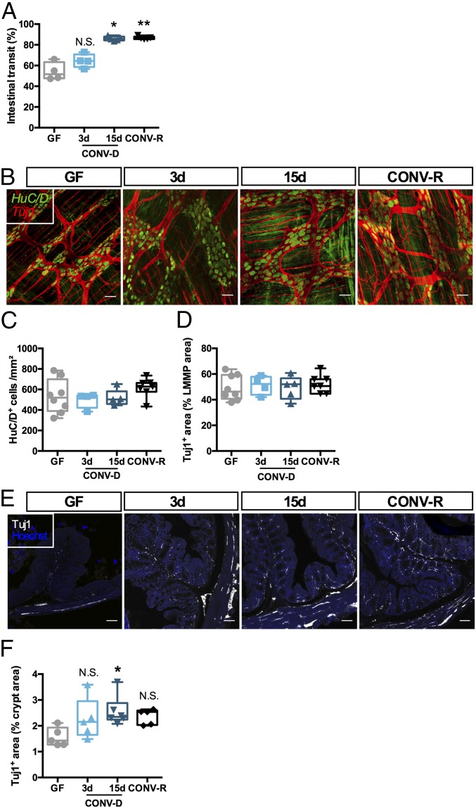
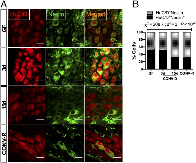
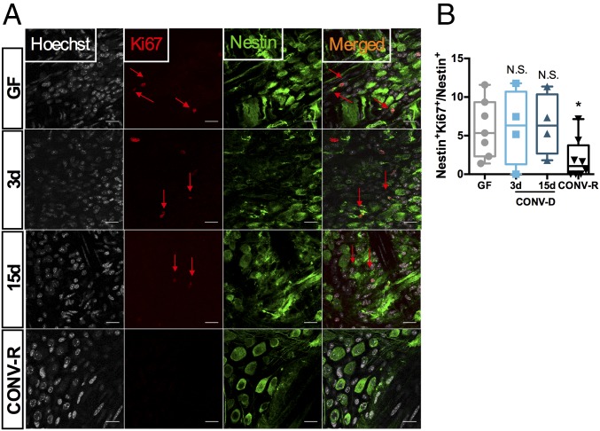
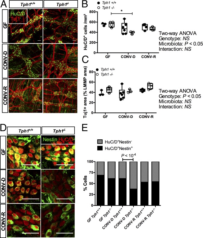
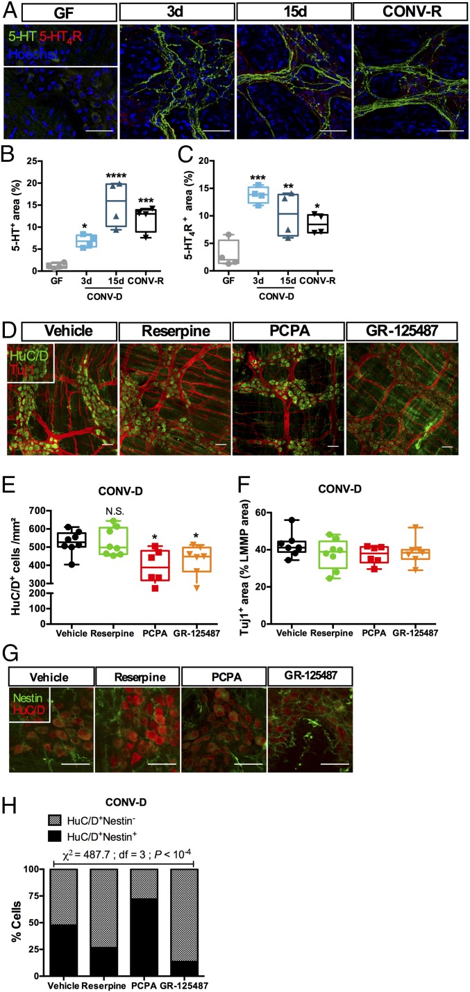
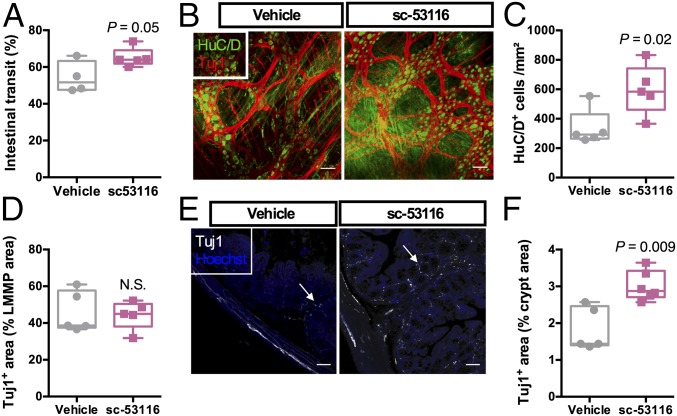
The enteric nervous system (ENS) is crucial for essential gastrointestinal physiologic functions such as motility, fluid secretion, and blood flow. The gut is colonized by trillions of bacteria that regulate host production of several signaling molecules including serotonin (5-HT) and other hormones and neurotransmitters. Approximately 90% of 5-HT originates from the intestine, and activation of the 5-HT4 receptor in the ENS has been linked to adult neurogenesis and neuroprotection. Here, we tested the hypothesis that the gut microbiota could induce maturation of the adult ENS through release of 5-HT and activation of 5-HT4 receptors. Colonization of germ-free mice with a microbiota from conventionally raised mice modified the neuroanatomy of the ENS and increased intestinal transit rates, which was associated with neuronal and mucosal 5-HT production and the proliferation of enteric neuronal progenitors in the adult intestine. Pharmacological modulation of the 5-HT4 receptor, as well as depletion of endogenous 5-HT, identified a mechanistic link between the gut microbiota and maturation of the adult ENS through the release of 5-HT and activation of the 5-HT4 receptor. Taken together, these findings show that the microbiota modulates the anatomy of the adult ENS in a 5-HT-dependent fashion with concomitant changes in intestinal transit.

Keywords: 5-HT4R; enteric nervous system; microbiota; serotonin.


Conflict of interest statement

Conflict of interest statement: F.B. is cofounder of and shareholder in Metabogen AB.

Figures

Fig. 1.
Fig. 1.
The gut microbiota regulates ENS anatomy and function. (A) Intestinal transit in GF mice, GF mice colonized (CONV-D) with microbiota from a CONV-R donor for 3 or 15 d, and CONV-R mice. *P < 0.05; **P < 0.01 vs. GF; N.S., not significant; one-way ANOVA followed by Dunnett’s post hoc test. (B) Representative images of the LMMP of the colon showing pan-neuronal marker HuC/D (green) and neuron-specific beta-III tubulin (Tuj1, red). (C and D) Quantification of HuC/D+ cells (C) and the Tuj1+ area (D). (E and F) Representative images (E) and quantification (F) of the innervation of the colonic crypts of the mice using the peripheral neuronal marker Tuj1. *P < 0.05 vs. GF; N.S., not significant; Kruskal–Wallis test followed by Dunn’s post hoc test. (Scale bars: 50 µm.)
Fig. 2.
Fig. 2.
Colonization of GF mice with a gut microbiota induces maturation of neuronal precursors in the myenteric plexus of the colon. (A) Immunostaining with HuC/D (red) and the neuronal precursor marker Nestin (green). (B) Colonization reduced the proportion of Nestin+ neurons in the myenteric ganglia (n > 1,000 cells counted per group). (Scale bars: 20 µm.)
Fig. 3.
Fig. 3.
Colonization of adult GF mice with a gut microbiota results in cycling of neuronal progenitors. (A) Representative images of a colonic myenteric ganglion of a mouse stained with the cycling cell marker Ki67 (red arrows), neuronal precursor marker Nestin (green), and with nuclei counterstained with Hoechst (gray). (B) Quantification of double-positive Nestin/Ki67 cells. *P < 0.05 vs. GF; N.S., not significant; Kruskal–Wallis test followed by Dunn’s post hoc test. (Scale bars: 20 µm.)
Fig. 4.
Fig. 4.
Mucosal 5-HT is neuroprotective in CONV-D mice. (A) Representative images of the colonic LMMP showing pan-neuronal marker HuC/D (green) and neuron-specific β-III tubulin (Tuj1, red). (B and C) Quantification of HuC/D+ cells (B) and the Tuj1+ area (C). P values are reported after two-way ANOVA followed by Dunnett’s multiple comparisons test. *P < 0.05. (D) Immunostaining with HuC/D (red) and the neuronal precursor marker Nestin (green). (E) Quantification of results in D. Split panels are provided in SI Appendix, Fig. S4E. P values reported are knockout vs. wild type; Fisher’s exact test (n > 750 cells were counted per group). (Scale bars: 50 µm.)
Fig. 5.
Fig. 5.
The gut microbiota regulates neuronal 5-HT release and 5-HT4R activation in the myenteric plexus. (A) Representative images of the colonic myenteric plexus showing immunoreactivity to 5-HT (green) and 5-HT4R (red). Localization of 5-HT and 5-HT4R was altered by colonization. (B and C) Quantification of immunoreactivity to 5-HT (B) and 5-HT4R (C). *P < 0.05; **P < 0.01; ***P < 0.001; ****P < 10−4 vs. GF; Kruskal–Wallis test followed by Dunn’s post hoc test. (D) Representative images of the colonic LMMP of CONV-D mice treated with a vehicle solution, the 5-HT4R antagonist GR-125487, the TPH inhibitor PCPA, or the vesicular monoamine transporter blocker reserpine, showing the pan-neuronal marker HuC/D (green) and neuron-specific β-III tubulin (Tuj1, red). (E and F) Quantification of HuC/D+ cells (E) and the Tuj1+ area (F). *P < 0.05 vs. GF; N.S., not significant; Kruskal–Wallis test followed by Dunn’s post hoc test. (G) Immunostaining of HuC/D (red) and the neuronal precursor marker Nestin (green). Split panels are shown in SI Appendix, Fig. S6. (H) Quantification of the proportion of Nestin+ neurons (n > 500 cells counted per group). (Scale bars: 50 µm.)
Fig. 6.
Fig. 6.
5-HT4R regulates ENS anatomy and function in GF mice. (A) Intestinal transit in GF mice that were given the 5-HT4R antagonist sc-53116 or a vehicle solution. P = 0.05; Student’s unpaired t test. (B) Representative images of the colonic LMMP of the aforementioned mice showing the pan-neuronal marker HuC/D (green) and neuron-specific β-III tubulin (Tuj1, red). (C and D) Quantification of HuC/D+ cells (C) and the Tuj1+ area (D). (E) Representative images of the innervation of the colonic crypts of the mice (white arrows) using the peripheral neuronal marker Tuj1. (F) Quantification of the Tuj1+ area. P values were determined by the Mann–Whitney test. N.S., not significant. (Scale bars: 50 µm.)

Comment in

  • Microbiota modulate ENS maturation.
    Dickson I. Dickson I. Nat Rev Gastroenterol Hepatol. 2018 Aug;15(8):454-455. doi: 10.1038/s41575-018-0040-7. Nat Rev Gastroenterol Hepatol. 2018. PMID: 29934558 No abstract available.

References

    1. Furness JB. The enteric nervous system and neurogastroenterology. Nat Rev Gastroenterol Hepatol. 2012;9:286–294. - PubMed
    1. Joseph NM, et al. Enteric glia are multipotent in culture but primarily form glia in the adult rodent gut. J Clin Invest. 2011;121:3398–3411. - PMC - PubMed
    1. Laranjeira C, et al. Glial cells in the mouse enteric nervous system can undergo neurogenesis in response to injury. J Clin Invest. 2011;121:3412–3424. - PMC - PubMed
    1. Liu M-T, Kuan Y-H, Wang J, Hen R, Gershon MD. 5-HT4 receptor-mediated neuroprotection and neurogenesis in the enteric nervous system of adult mice. J Neurosci. 2009;29:9683–9699. - PMC - PubMed
    1. Pham TD, Gershon MD, Rothman TP. Time of origin of neurons in the murine enteric nervous system: Sequence in relation to phenotype. J Comp Neurol. 1991;314:789–798. - PubMed

Publication types