. 2018 Jun 19;115(25):E5796-E5804.
doi: 10.1073/pnas.1707702115. Epub 2018 Jun 4.

Stimulation-induced increases in cerebral blood flow and local capillary vasoconstriction depend on conducted vascular responses

Affiliations

Stimulation-induced increases in cerebral blood flow and local capillary vasoconstriction depend on conducted vascular responses

Changsi Cai et al. Proc Natl Acad Sci U S A. .

Abstract

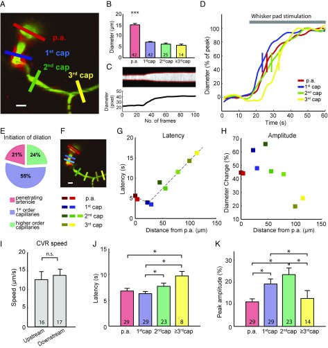
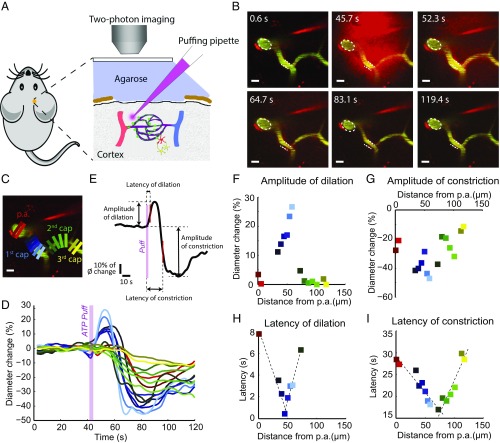
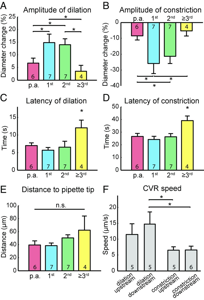
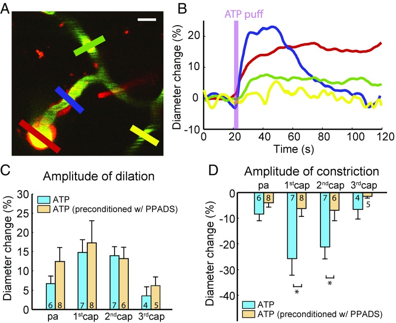
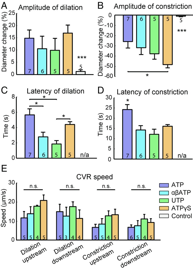
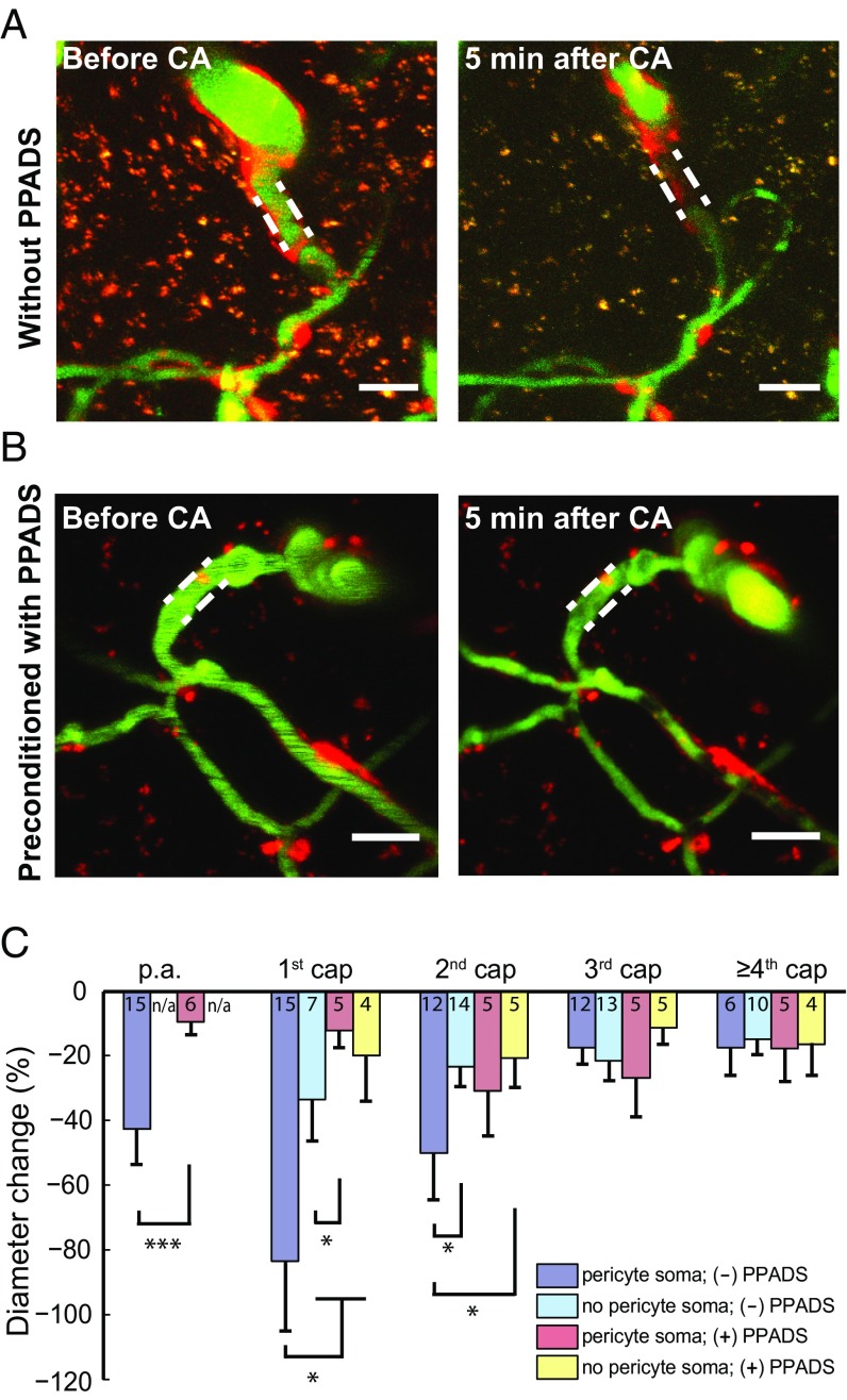
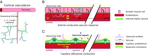
Functional neuroimaging, such as fMRI, is based on coupling neuronal activity and accompanying changes in cerebral blood flow (CBF) and metabolism. However, the relationship between CBF and events at the level of the penetrating arterioles and capillaries is not well established. Recent findings suggest an active role of capillaries in CBF control, and pericytes on capillaries may be major regulators of CBF and initiators of functional imaging signals. Here, using two-photon microscopy of brains in living mice, we demonstrate that stimulation-evoked increases in synaptic activity in the mouse somatosensory cortex evokes capillary dilation starting mostly at the first- or second-order capillary, propagating upstream and downstream at 5-20 µm/s. Therefore, our data support an active role of pericytes in cerebrovascular control. The gliotransmitter ATP applied to first- and second-order capillaries by micropipette puffing induced dilation, followed by constriction, which also propagated at 5-20 µm/s. ATP-induced capillary constriction was blocked by purinergic P2 receptors. Thus, conducted vascular responses in capillaries may be a previously unidentified modulator of cerebrovascular function and functional neuroimaging signals.

Keywords: cerebral capillaries; conducted vascular responses; neurovascular coupling; pericytes; purinergic signaling.


Conflict of interest statement

The authors declare no conflict of interest.

Figures

Fig. 1.
Fig. 1.
Functional vessel dilation in the mouse barrel cortex. (A) A two-photon image of the barrel cortex of a NG2-DsRed mouse at ∼150 µm depth. The p.a.s branch out a capillary horizontally (first order). Further branches are defined as second- and third-order capillaries. Pericytes are labeled with a red fluorophore (NG2-DsRed) and the vessel lumen with FITC-dextran (green). ROIs are placed across the vessel to allow measurement of the vessel diameter (colored bars). (Scale bar: 10 µm.) (B) Vessel diameters at different orders of capillaries. p.a., 15.09 ± 4.15 μm; 1st cap (first-order capillaries), 7.18 ± 1.93 μm; 2nd cap (second-order capillaries), 6.25 ± 2.43 μm; 3rd cap (third-order capillaries), 7.63 ± 2.47 μm. The p.a. diameter is significantly larger than all orders of capillaries. ***P < 0.001, one-way ANOVA with post hoc test. (C) Example trace of fluorescent intensity over time at the blue ROI in A is shown as the gray image, and the two red curves indicate the vessel wall (Upper). The distance of the two red curves is calculated as the time course of vessel diameter (Lower). (D) The normalized diameter change over time at different orders of capillaries in response to whisker-pad stimulation. The short vertical bar is where the curve reaches 50% peak, which is defined as response onset. (E) Distribution of the locations where the functional dilation initiated (n = 29 locations). (F) Multiple ROIs at the p.a. and first-, second-, and third-order capillaries are marked as red, blue, green, and yellow, respectively. (Scale bar: 10 µm.) (G) In this mouse experiment, the half-maximal dilation latency of each ROI is plotted with corresponding colors on the left along the geographic distance from the p.a. Dashed lines show the linear fit of the conducted dilation. (H) The maximal dilation amplitude is plotted with corresponding colors on the left along the geographic distance from the p.a. (I) Eighteen out of 29 imaged vasculatures exhibited conducted functional dilation, with an upstream conductive speed of 12.65 ± 0.96 µm/s and downstream conductive speed of 12.83 ± 0.64 µm/s. No significant difference was found between upstream and downstream conductive speeds. n.s., not significant; P > 0.05, unpaired t test. (J) Time to 50% maximal dilation was significantly longer in the third-order capillaries than the p.a. and first-order capillaries. The second-order capillaries dilated significantly slower than the first-order capillaries. *P < 0.05, one-way ANOVA with post hoc test. (K) Maximal dilation amplitude in different order capillaries. First- and second-order capillaries exhibited significantly larger responses than other locations. *P < 0.05, one-way ANOVA with post hoc test. All error bars represent SEM.
Fig. 2.
Fig. 2.
ATP puffing by micropipette induces vessel dilation, followed by constriction. (A) Diagram of the in vivo experimental setup. The puffing micropipette is placed in proximity of the near-arteriole site. The micropipette contains a mixture of 10 µM Alexa 594 (red color in glass micropipette) and 1 mM ATP. (B) Video snapshots of the time course of puffing with 1 mM ATP from the micropipette. Vessel dilation precedes constriction. Dashed lines indicate the vessel contours at the resting state. (Scale bars: 10 µm.) (C) Multiple uniquely colored ROIs are placed along the vasculature to measure the vessel diameter. (Scale bar: 10 µm.) (D) Normalized diameter change is plotted over time for each ROI. The ROIs and trace color are coded identically. (E) Amplitudes of dilation or constriction are defined as positive or negative amplitudes at the maximal vascular response. The latencies of dilation and constriction are reported as time to half positive or negative maximum after puffing onset. (FI) In this mouse experiment, the distribution of all ROIs in C and D with amplitude of dilation (F), amplitude of constriction (G), latency of dilation (H), and latency of constriction (I) over the geographic distance from the p.a. along the vasculature. The dashed lines represent the linear fit of the upstream and downstream conductive responses.
Fig. 3.
Fig. 3.
ATP-puffing-induced dilation and constriction vary in amplitude and latency at different-order capillaries. (A) The amplitudes of dilation and (B) constriction are significantly higher at first- and second-order capillaries compared with other order capillaries. (C) The latencies of dilation and (D) constriction are significantly longer for third-order and higher capillaries than other order capillaries. *P < 0.05, one-way ANOVA with post hoc test. (E) The mean distance from the pipette tip to the vessels of the different branch orders. n.s., not significant; P > 0.05, one-way ANOVA with post hoc test. (F) Comparison of upstream and downstream conductive speeds of ATP-puffing-induced dilation and constriction. *P < 0.05, one-way ANOVA with post hoc test. All error bars represent SEM.
Fig. 4.
Fig. 4.
ATP-puffing-induced constriction is mediated by purinergic type 2 receptors. (A) After topical application of 0.5 mM PPADS, the puffing pipette is placed in proximity of a near-arteriole site. Red, blue, green, and yellow ROIs are placed at the p.a. and first-, second-, and third-order capillaries to measure the diameter, respectively. (Scale bar: 10 µm.) (B) Time course of the diameter change in each ROI indicated in A. Preconditioning with 0.5 mM PPADS and puffing with 1 mM ATP induces dilation but profoundly attenuates constriction. (C) The amplitude of dilation is not significantly different with and without the application of PPADS among all orders of capillaries. n.s., not significant; P > 0.05, unpaired t test. (D) The amplitude of constriction is significantly different with and without preconditioning with PPADS on the first- and second-order capillaries. *P < 0.05, unpaired t test. All error bars represent SEM.
Fig. 5.
Fig. 5.
Vessel responses of first-order capillaries to puffing with ATP, P2X, P2Y receptor agonists, ATPγS, and red dye. (A) Comparison of different puffing compounds with amplitude of dilation, (B) amplitude of constriction, (C) latency of dilation, (D) latency of constriction, and (E) conductive speed. The compounds are 1 mM ATP, 1 mM P2X receptor agonist (αβATP), 1 mM P2Y receptor agonist (UTP), 1 mM ATPγS, and 10 µM Alexa 594 as control. n/a, not available; *P < 0.05, ***P < 0.001, one-way ANOVA with post hoc test. Note that the latency of the control experiment is marked as not available for both dilation (C) and constriction (D). This is due to the small responses upon control puffing and the suboptimal measurements of latency. All error bars represent SEM. n.s., not significant.
Fig. 6.
Fig. 6.
Ischemia leads to severe constriction of capillaries at the near-arteriole site and preconditioning of P2 receptor blockers mitigates constriction of capillaries. (A) Image stacks (1-µm step size, average intensity projection) of the vasculature, including the p.a. and first few orders of capillaries. Five minutes after ischemia by cardiac arrest (CA), severe constriction was observed at the p.a. and first- and second-order capillaries, but third-order and higher capillaries were moderately constricted. Dashed lines indicate the vessel contours of first-order capillaries before cardiac arrest. (Scale bars: 20 µm.) (B) Preconditioning with 0.5 mM PPADS for 2 h rescued severe constriction of the p.a. and first-order capillary 5 min after CA. Dashed lines indicate the vessel contours of first-order capillaries before CA. (Scale bars: 20 µm.) (C) The most severe constrictions at the first- and second-order capillaries colocalized with pericytes. The third-order and higher capillaries exhibited moderate constriction. Preconditioning with PPADS mitigated vasoconstriction at the p.a. and first- and second-order capillaries. For the p.a., an unpaired t test was used. n/a, not available; ***P < 0.001. For the other order capillaries, one-way ANOVA with post hoc test was used (*P < 0.05). All error bars represent SEM.
Fig. 7.
Fig. 7.
Possible mechanisms of ATP-puffing-induced dilation and constriction. (A) The pial artery and p.a.s consist of endothelium surrounded by smooth muscle cells (light red). As capillaries branch off the p.a., smooth muscle is replaced by pericytes (light blue) with heterogeneous morphologies across first-, second-, and higher-order capillaries (i.e., going from the p.a. to the venous side). (B) Fast and long-range conduction along arterioles and arteries via electrical conduction and the local Ca2+ wave. (C) Observed slow and low-range conduction of vasomotor responses emanating mostly from first- and second-order capillaries seem to involve signaling by diffusion. Both paracrine signaling along astrocyte end-feet and intracellular diffusion along putative gap junctions can be envisioned.

References

    1. Abbott NJ, Patabendige AA, Dolman DE, Yusof SR, Begley DJ. Structure and function of the blood-brain barrier. Neurobiol Dis. 2010;37:13–25. - PubMed
    1. Sakadžić S, et al. Large arteriolar component of oxygen delivery implies a safe margin of oxygen supply to cerebral tissue. Nat Commun. 2014;5:5734. - PMC - PubMed
    1. Hall CN, et al. Capillary pericytes regulate cerebral blood flow in health and disease. Nature. 2014;508:55–60. - PMC - PubMed
    1. Gould IG, Tsai P, Kleinfeld D, Linninger A. The capillary bed offers the largest hemodynamic resistance to the cortical blood supply. J Cereb Blood Flow Metab. 2017;37:52–68. - PMC - PubMed
    1. Peppiatt CM, Howarth C, Mobbs P, Attwell D. Bidirectional control of CNS capillary diameter by pericytes. Nature. 2006;443:700–704. - PMC - PubMed

Publication types

MeSH terms

Substances