. 2018 Jul 4;145(13):dev164855.
doi: 10.1242/dev.164855.

Sox30 initiates transcription of haploid genes during late meiosis and spermiogenesis in mouse testes

Affiliations

Sox30 initiates transcription of haploid genes during late meiosis and spermiogenesis in mouse testes

Shun Bai et al. Development. .

Erratum in

Abstract

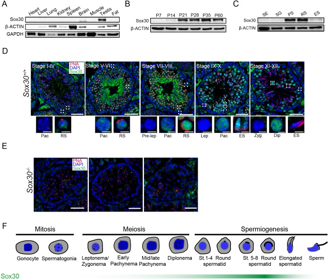
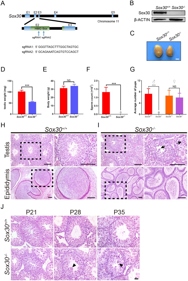
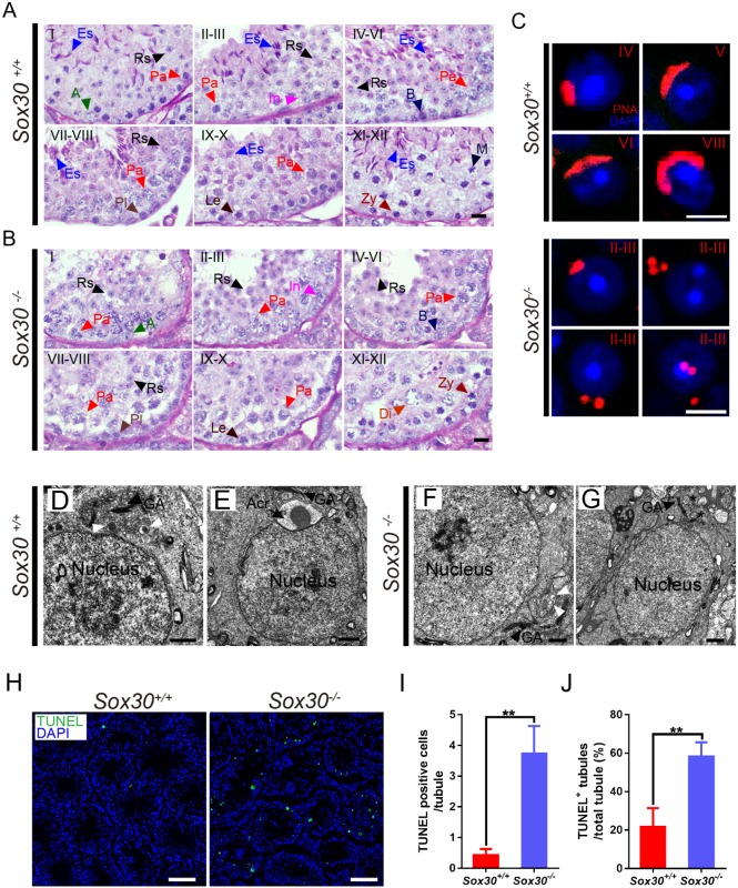
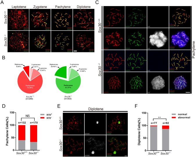
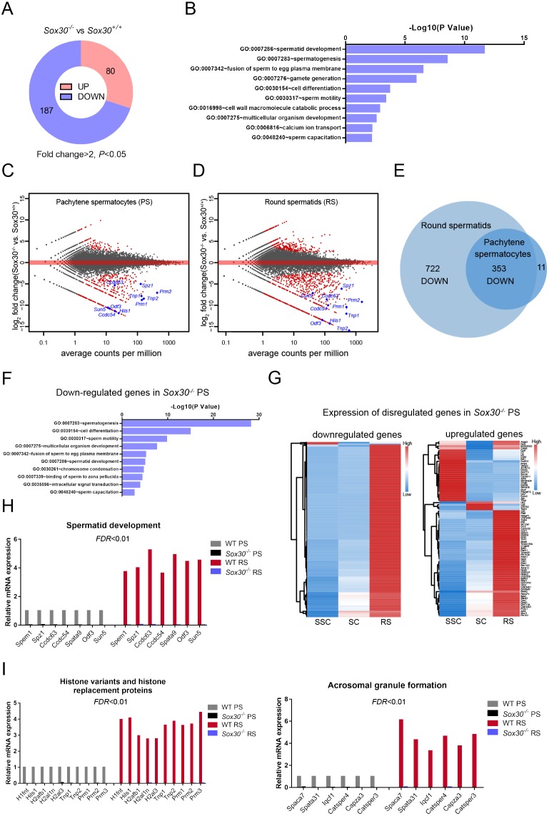
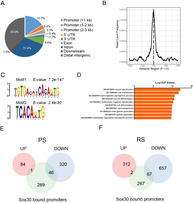
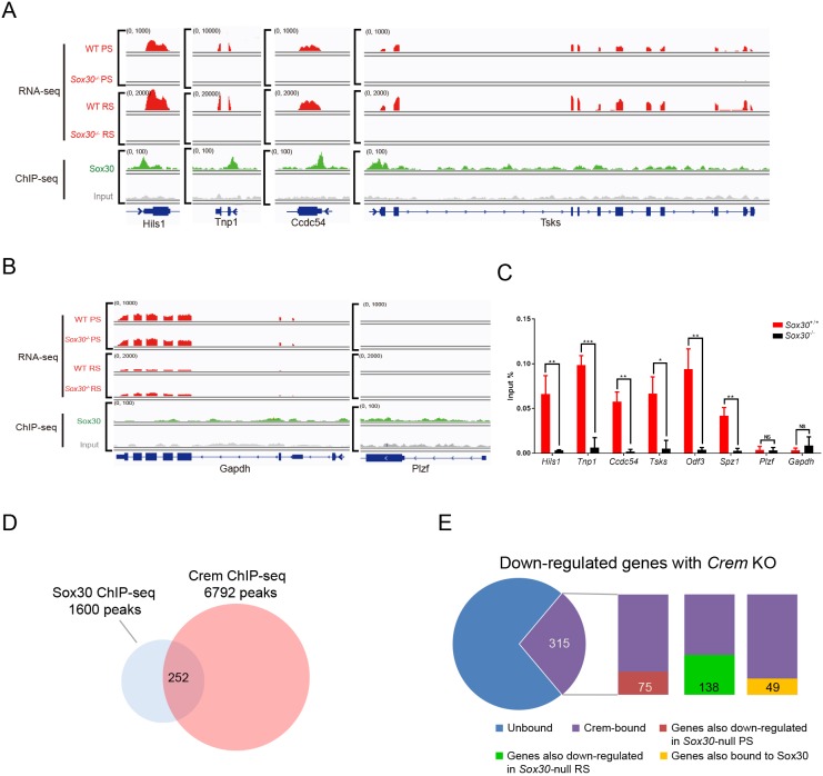
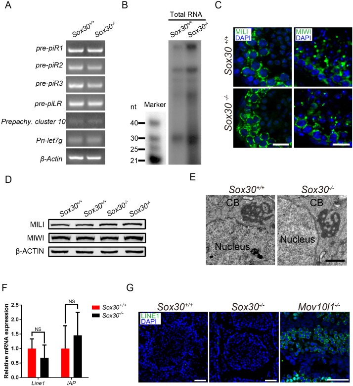
Transcription factors of the Sox protein family contain a DNA-binding HMG box and are key regulators of progenitor cell fate. Here, we report that expression of Sox30 is restricted to meiotic spermatocytes and postmeiotic haploids. Sox30 mutant males are sterile owing to spermiogenic arrest at the early round spermatid stage. Specifically, in the absence of Sox30, proacrosomic vesicles fail to form a single acrosomal organelle, and spermatids arrest at step 2-3. Although most Sox30 mutant spermatocytes progress through meiosis, accumulation of diplotene spermatocytes indicates a delayed or impaired transition from meiotic to postmeiotic stages. Transcriptome analysis of isolated stage-specific spermatogenic cells reveals that Sox30 controls a core postmeiotic gene expression program that initiates as early as the late meiotic cell stage. ChIP-seq analysis shows that Sox30 binds to specific DNA sequences in mouse testes, and its genomic occupancy correlates positively with expression of many postmeiotic genes including Tnp1, Hils1, Ccdc54 and Tsks These results define Sox30 as a crucial transcription factor that controls the transition from a late meiotic to a postmeiotic gene expression program and subsequent round spermatid development.

Keywords: Gene regulation; Male germ cell; Mouse; Sox30; Spermiogenesis.


Conflict of interest statement

Competing interestsThe authors declare no competing or financial interests.

Figures

Fig. 1.
Fig. 1.
Sox30 is highly enriched in mouse testes. (A-C) Western blot analysis of Sox30 protein in lysates from adult mouse tissues (A), from mouse testis tissue collected at different time points during postnatal development (B) and from isolated spermatogenic cell populations, including Sertoli cells (SE), spermatogonia (SG), pachytene spermatocytes (PS), round spermatids (RS) and elongating spermatids (ES). β-actin serves as an internal loading control. (D,E) Immunostaining of testis sections from 8-week-old wild-type (D) and Sox30−/− mutant (E) mice for Sox30 (green) and PNA (red). PNA is an acrosome marker. DNA was counterstained with DAPI. Lower panels show magnifications of the boxed area in the upper panels. Dip, diplotene; ES, elongating spermatids; Pac, pachytene; pre-lep, pre-leptotene; RS, round spermatids; Zyg, zygotene. Scale bars: 50 μm. (F) Graphic representation of Sox30 expression (green bar) during spermatogenesis.
Fig. 2.
Fig. 2.
Sox30 mutant mice arrest at the round spermatid stage. (A) Diagram illustrating the CRISPR/Cas9 targeting strategy, including position and sequence of guide RNAs (sgRNAs). E, exon. (B) Western blot analysis confirms absence of Sox30 protein in lysates from 8-week-old Sox30−/− mutant mice. β-actin serves as an internal loading control. (C) Morphological appearance of testis from Sox30−/−mice versus Sox30+/+ (wild-type) mice revealing smaller size. (D-F) Comparison of testis weight (D), body weight (E) and epididymal sperm counts (F) of 8-week-old Sox30+/+ and Sox30−/− mice. Data are presented as mean±s.d. (D,E: n=6 per group; F: n=5) ***P<0.001; NS, not significant (Student's t-test). (G) Fertility test for adult Sox30−/− males and Sox30−/− females. Each genotype shown was coupled with fertile wild-type mates. n=3 for each genotype. ***P<0.001; NS, not significant (Student's t-test). (H,I) Hematoxylin and Eosin staining of testis tubules from 8-week-old wild-type (H) and Sox30−/− (I) mice. Sox30−/− tubules were devoid of elongating spermatids. Black arrows mark multinucleated cells. Right-hand panels show magnifications of the boxed areas on the left. (J) Sox30+/+ (wild-type) epididymal tubules were full of spermatozoa whereas epididymal tubules from Sox30−/− mice were devoid of mature sperm but contained degenerating cells with round-shaped nuclei (black arrowheads). Scale bars: 100 μm.
Fig. 3.
Fig. 3.
Sox30-deficient round spermatids exhibit impaired fusion of proacrosomic vesicles and arrest at step 2-3. (A,B) PAS- and Hematoxylin-stained testis sections from 8-week-old Sox30+/+ (A) and Sox30−/− (B) mice. The seminiferous tubule stage (top left) was identified based on the PAS staining pattern and arrangement of spermatogenic cells. A, type A spermatogonia; B, type B spermatogonia; Es, elongating spermatids; In, intermediate spermatogonia; Le, leptotene spermatocytes; M, meiotic division; Pa, pachytene spermatocytes; Pl, preleptotene spermatocytes; Rs, round spermatids; Zy, zygotene spermatocytes. Scale bars: 50 μm. (C) Fluorescently tagged PNA-staining (red) of testis sections from 8-week-old mice shows acrosome formation from proacrosomic vesicles in step 4-8 wild-type spermatids but failure of vesicle fusion in cells from Sox30−/− mice. DNA was counterstained with DAPI. Scale bars: 10 μm. (D-G) Electron microscopic analysis of testis tissue from 8-week-old Sox30+/+ (D,E) and Sox30−/− (F,G) mice. Step 2-3 wild-type round spermatids contain proacrosomal vesicles (white arrowheads) located between the Golgi apparatus and the nucleus (D), and a newly assembled acrosome is visible in a step 4 round spermatid (E). (F,G) Sox30−/− spermatids at step 2-3 contain proacrosomic vesicles, but a large acrosome was never observed. A subset of proacrosomic vesicles were not located between the Golgi apparatus and the nucleus (G). White arrowheads mark proacrosomal vesicles; asterisks identify proacrosomal vesicles with abnormal localization. Acr, acrosome; GA, Golgi apparatus. Scale bars: 1 μm. (H-J) Increased apoptosis in Sox30−/− testis. (H) TUNEL assay in testis sections from 8-week-old Sox30+/+ and Sox30−/− males. Scale bars: 100 μm. (I) Quantification of TUNEL-positive cells per tubule. n=3 for each genotype. (J) Percentage of TUNEL-positive tubules in testis from 8-week-old Sox30+/+ and Sox30−/− mice. Tubules examined: Sox30+/+, n=96, Sox30−/−, n=156. Data presented are mean±s.d. **P<0.01 (Student's t-test).
Fig. 4.
Fig. 4.
Increased proportion of diplotene spermatocytes in Sox30−/− testis. (A,B) Immunolabeling of spermatocyte spread nuclei from 8-week-old wild-type and Sox30−/− mice was performed using antibodies against SYCP1 (green) and SYCP3 (red), which are the transverse and lateral elements of synaptonemal complex that forms on the chromosome axis. Differential stages of meiotic I prophase I (leptotene, zygotene, pachytene and diplotene) were identified based on the staining pattern of SYCP1 and SYCP3. At the leptotene stage, axis elements are not completely formed; SYCP3 staining appears as short and discrete lines in the nucleus and SYCP1 signal is absent (Yuan et al., 2000; Yang et al., 2006). During the subsequent zygotene stage, homologous chromosomes start to pair and are associated via the transverse element SYCP1. Axis elements become thicker and SYCP1 is detected at the regions where chromosomes pair. At pachytene, autosomes fully synapse except for the X and Y chromosomes, which only synapse at a short pseudoautosomal region (PAR). SYCP1 and SYCP3 co-localized at autosomes, and the SYCP1 signal is detected in the short region where XY chromosomes synapse (Zickler and Kleckner, 1999; Perry et al., 2001). At diplotene, synapsed chromosomes start to separate from each other and the lateral elements become disassociated in certain regions, where SYCP1 signal disappears. (B) Percentage of spermatocytes at each stage of meiotic prophase I (leptonema, zygonema, pachynema and diplonema) in 8-week-old Sox30+/+ and Sox30−/− mice (spermatocytes analyzed: Sox30+/+, n=249; Sox30−/−, n=353). (C,D) Triple immunostaining for SYCP3 (red), SYCP1 (green), and H1t (white) reveals a normal proportion of early (H1t) and mid-to-late (H1t+) pachytene spermatocytes in 8-week-old Sox30−/− mice. NS, not significant (χ2 test). (E) Double immunostaining for γH2AX (green) and SYCP3 (red) identifies abnormal localization of γH2AX in diplotene spermatocytes from 8-week-old Sox30−/− mice. (F) Quantification analysis for diplotene spermatocytes with abnormal γH2AX distribution. Sox30+/+, n=77; Sox30−/−, n=92. **P<0.01 (χ2 test). Scale bars: 10 μm.
Fig. 5.
Fig. 5.
Sox30−/− deficiency produces global and cell type-specific gene expression changes in the testis. (A) Number of genes exhibiting significant (fold change >2, P<0.05) up- or downregulation in Sox30−/− versus Sox30+/+ testis at P21. n=3 for each genotype, each of which were deep sequenced separately. (B) Gene ontology associations of downregulated genes in Sox30−/− in testis identified by RNA-seq. (C,D) Scatter plot of differentially expressed transcripts in Sox30−/− pachytene spermatocytes (C) and round spermatids (D) compared with Sox30+/+ cells. Pachytene spermatocytes and round spermatids were isolated from 8-week-old wild-type and Sox30−/− mice by the STA-PUT method. Wild-type, n=6; Sox30−/−, n=8. Each red dot represents a gene that was significantly changed (fold change >2, FDR<0.01). (E) Analysis of differential gene expression in Sox30−/− pachytene spermatocytes and round spermatids identifies 353 transcripts that are downregulated in both stages. (F) GO term enrichment analysis for downregulated transcripts in pachytene spermatocytes of Sox30−/− mice. (G) Genes upregulated or downregulated in Sox30−/− pachytene spermatocytes were used to produce a heat map exhibiting their expression pattern in stage-specific spermatogenic cells. RS, round spermatids; SC, pachytene spermatocytes; SSC, spermatogonial stem cells. (H,I) Expression changes in genes encoding factors essential for spermatid development (H), histone variants and replacement associated proteins (I) and factors involved in acrosomal granule formation (I).
Fig. 6.
Fig. 6.
ChIP-seq analysis revealed Sox30-binding sites in mouse testis. ChIP-seq experiments with anti-Sox30 antibody were performed using wild-type mouse testes at P28, when Sox30 is abundantly expressed. (A) Genomic locations of Sox30 binding sites. (B) Distribution of Sox30 ChIP reads on gene bodies is plotted. Sox30 was enriched at the regions from −1 kb to +1 kb relative to the TSS. The y-axis represents the frequency of Sox30 ChIP-seq counts at a specific site, normalized by total read counts. (C) De novo motif analysis of Sox30-binding sites using MEME. The most enriched de novo motifs are shown (Motif 1: E value=7.2e−147; Motif 2: E value=2.4e−30). (D) The gene ontology terms analyzed from the top 50% of Sox30-bound genes. (E,F) Identification of direct target genes of Sox30 by ChIP-seq and RNA-seq analysis. The total number of genes with Sox30 ChIP-seq peaks at promoters was compared with disregulated genes upon Sox30 deletion in pachytene spermatocytes (E) and round spermatids (F).
Fig. 7.
Fig. 7.
Sox30 specifically bound and directly regulated expression of postmeiotic genes. (A,B) Genome browser view of Sox30 ChIP-seq and RNA-seq reads on representative gene loci in testes or isolated cells from wild-type and Sox30−/− mice. (C) ChIP-qPCR analysis of Sox30 enrichment on promoter regions of Hils1, Tnp1, Ccdc54, Tsks, Odf3, Spz1, Plzf and Gapdh in P28 testes of wild-type and Sox30−/− mice. The chromatin occupancy at Plzf and the housekeeping gene Gapdh was low and was similar in wild type and mutant. Each sample represents a pool of three individual mice. Data are presented as mean±s.d. *P<0.05, **P<0.01, ***P<0.001 (Student's t-test). (D) Overlapping binding sites of Sox30 and CREM ChIP-seq peaks in testis. (E) Out of a total of 820 genes downregulated in Crem−/− testis, 315 genes contained CREM binding peaks. Of those, 75 and 138 genes were also reduced in Sox30−/− pachytene spermatocytes and Sox30−/− pachytene spermatocytes, respectively. Of the 315 direct targets of CREM, 49 genes were bound by Sox30 at promoters or intragenic regions.
Fig. 8.
Fig. 8.
Sox30−/− deficiency does not affect transcription and processing of piRNA precursors in the testis. (A) Expression of piRNA precursors by semi-quantitative RT-PCR analysis in adult wild-type and mutant testes. (B) The level of piRNA abundance was not reduced in Sox30 mutant testes. 5′ labeling of total small RNAs in 8-week-old Sox30+/+ and Sox30−/− testis. (C) Immunostaining of the piRNA pathway components MILI and MIWI on frozen sections of adult Sox30+/+ and Sox30−/− testes. Scale bars: 10 μm. (D) Western blot analysis of MILI and MIWI in P21 wild-type and mutant testes. (E) Electron micrographs of wild-type and Sox30-deficient round spermatids. Scale bar: 1 μm. (F) Q-PCR analysis of LINE1 and IAP transcripts in P21 wild-type and mutant testes. NS, not significant (Student’s t-test). n=3. (G) Immunofluorescence detection of LINE1 ORF1p from adult wild-type, Sox30−/− and Mov10l1-deficent testes. Scale bars: 50 μm.

Similar articles

Cited by

References

    1. Barral S., Morozumi Y., Tanaka H., Montellier E., Govin J., de Dieuleveult M., Charbonnier G., Coute Y., Puthier D., Buchou T. et al. (2017). Histone variant H2A.L.2 guides transition protein-dependent protamine assembly in male germ cells. Mol. Cell 66, 89-101 e8. 10.1016/j.molcel.2017.02.025. - DOI - PubMed
    1. Bellvé A. R. (1993). Purification, culture, and fractionation of spermatogenic cells. Methods Enzymol. 225, 84-113. 10.1016/0076-6879(93)25009-Q - DOI - PubMed
    1. Bettegowda A. and Wilkinson M. F. (2010). Transcription and post-transcriptional regulation of spermatogenesis. Philos. Trans. R. Soc. Lond. B Biol. Sci. 365, 1637-1651. 10.1098/rstb.2009.0196 - DOI - PMC - PubMed
    1. Blendy J. A., Kaestner K. H., Weinbauer G. F., Nieschlag E. and Schütz G. (1996). Severe impairment of spermatogenesis in mice lacking the CREM gene. Nature 380, 162-165. 10.1038/380162a0 - DOI - PubMed
    1. Bolcun-Filas E., Bannister L. A., Barash A., Schimenti K. J., Hartford S. A., Eppig J. J., Handel M. A., Shen L. and Schimenti J. C. (2011). A-MYB (MYBL1) transcription factor is a master regulator of male meiosis. Development 138, 3319-3330. 10.1242/dev.067645 - DOI - PMC - PubMed

Publication types

Substances