. 2018 Aug 22;86(9):e00429-18.
doi: 10.1128/IAI.00429-18. Print 2018 Sep.

Methylthioadenosine Suppresses Salmonella Virulence

Affiliations

Methylthioadenosine Suppresses Salmonella Virulence

Jeffrey S Bourgeois et al. Infect Immun. .

Abstract

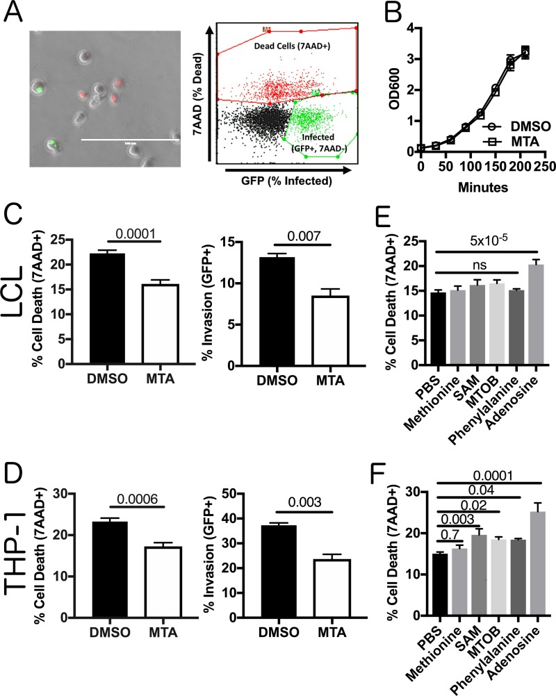
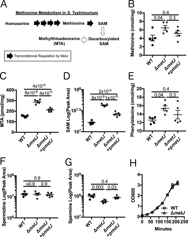
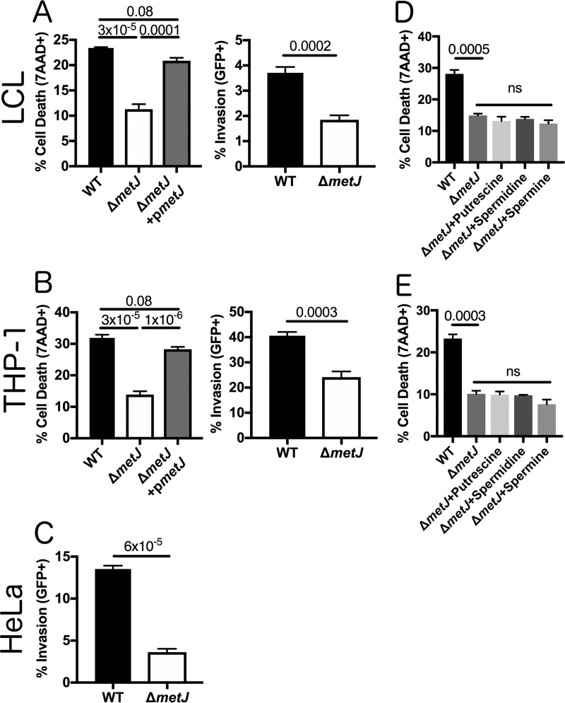
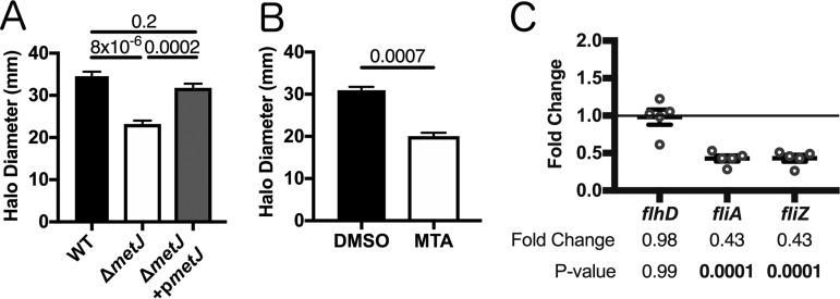
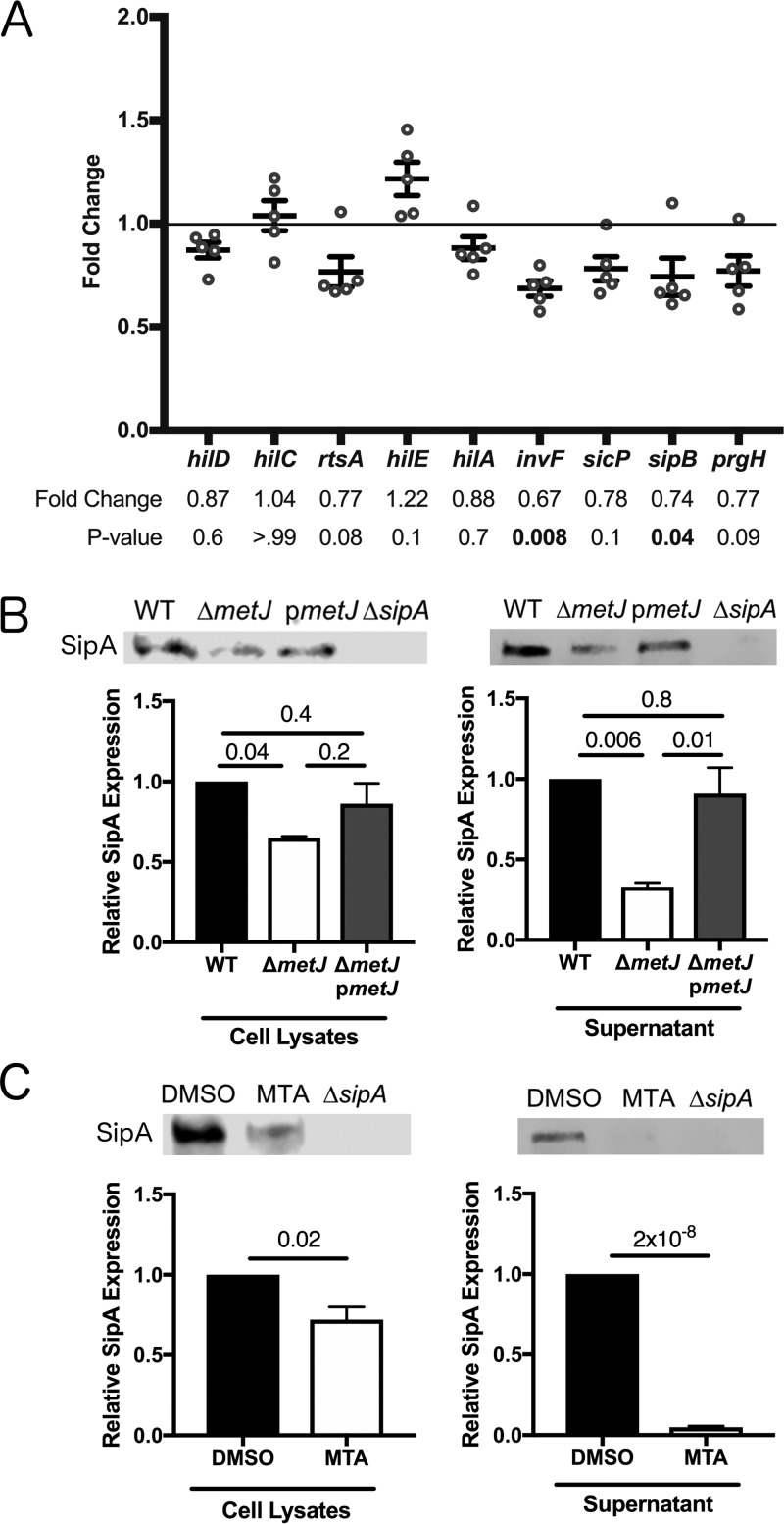
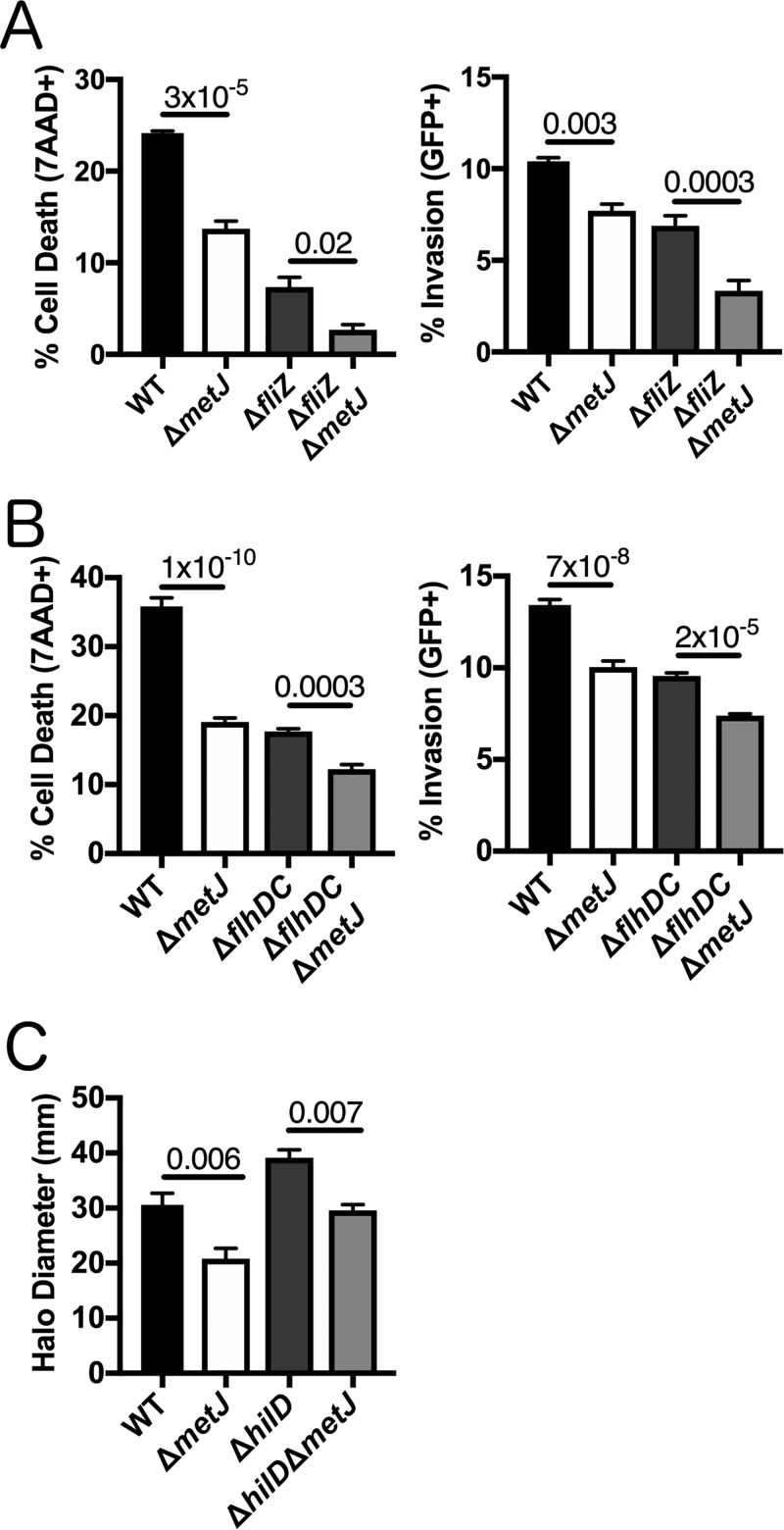
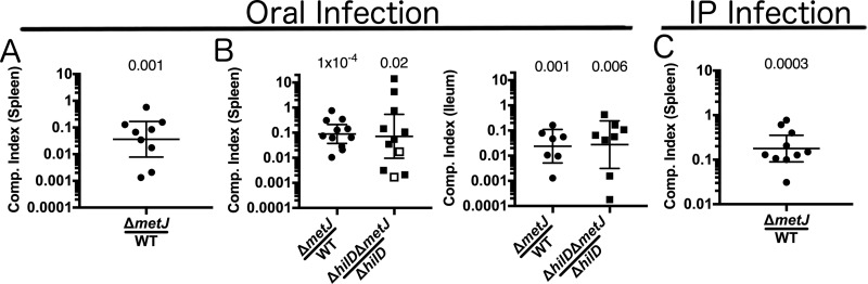
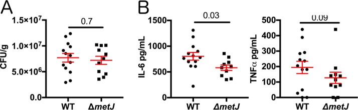
In order to deploy virulence factors at appropriate times and locations, microbes must rapidly sense and respond to various metabolite signals. Previously, we showed a transient elevation of the methionine-derived metabolite methylthioadenosine (MTA) concentration in serum during systemic Salmonella enterica serovar Typhimurium infection. Here we explored the functional consequences of increased MTA concentrations on S Typhimurium virulence. We found that MTA, but not other related metabolites involved in polyamine synthesis and methionine salvage, reduced motility, host cell pyroptosis, and cellular invasion. Further, we developed a genetic model of increased bacterial endogenous MTA production by knocking out the master repressor of the methionine regulon, metJ Like MTA-treated S Typhimurium, the ΔmetJ mutant displayed reduced motility, host cell pyroptosis, and invasion. These phenotypic effects of MTA correlated with suppression of flagellar and Salmonella pathogenicity island 1 (SPI-1) networks. S Typhimurium ΔmetJ had reduced virulence in oral and intraperitoneal infection of C57BL/6J mice independently of the effects of MTA on SPI-1. Finally, ΔmetJ bacteria induced a less severe inflammatory cytokine response in a mouse sepsis model. Together, these data indicate that exposure of S Typhimurium to MTA or disruption of the bacterial methionine metabolism pathway suppresses S Typhimurium virulence.

Keywords: SPI-1; Salmonella; flagellar motility; inflammation; metJ; metabolism; methionine salvage; methylthioadenosine; virulence regulation.


Figures

FIG 1
FIG 1
MTA treatment of S. Typhimurium reduces pyroptosis and invasion in vitro. (A) A modified gentamicin protection assay using an inducible GFP plasmid was used to detect pyroptosis (7-AAD+ red nuclear staining) and host cell invasion (intracellular GFP+ bacteria). This is observable by fluorescence microscopy and quantifiable by flow cytometry. Bar, 100 μm. (B) Exogenous MTA has no effect on the growth of S. Typhimurium in rich medium. The optical density at 600 nm (OD600) for S. Typhimurium treated with 300 μM MTA or 0.5% DMSO was measured every 30 min and showed that it exhibited equivalent growth with both treatments (n = 3). (C, D) Treatment of bacteria with 300 μM MTA during growth to late log phase (2 h 40 min) reduced pyroptosis (MOI, 30) and host cell invasion (MOI, 10) in LCLs measured at 3 h postinfection in 18592 LCLs (C) and THP-1 cells (D). Percent cell death represents all 7-AAD-positive cells under each infected condition, with the baseline uninfected cell death being subtracted. See the gating in panel A. Data were normalized to the global mean across five experiments, and P values, indicated at the top, were generated by a Student's t test. (E, F) Treating bacteria with other methionine-related metabolites (methionine, SAM, MTOB, and phenylalanine) or adenosine does not suppress host cell pyroptosis (MOI, 30), based on 3 to 5 independent experiments in 18592 LCLs (E) or THP-1 cells (F). Data were normalized to the global mean, and P values were generated by a one-way analysis of variance with Dunnett's multiple-comparison test. All error bars represent the standard error of the mean. ns, not significant.
FIG 2
FIG 2
metJ deletion results in disruption of methionine metabolism. (A) MetJ regulates the generation of methionine and SAM in S. Typhimurium by transcriptionally repressing the methionine regulon. Black arrows represent enzymes transcriptionally repressed by MetJ. (B to E) Deletion of metJ leads to increased methionine (B), MTA (C), SAM (D), and phenylalanine (E) levels, as measured by mass spectrometry (n = 5 biological replicates). (F, G) metJ deletion did not affect spermidine concentrations (F) but did result in decreased amounts of spermine (G). P values, indicated at the top, were generated through a one-way analysis of variance with Tukey's multiple-comparison test. (H) metJ deletion did not affect bacterial growth in LB (n = 3 biological replicates grown in LB plus 0.5% DMSO). All error bars represent the standard error of the mean.
FIG 3
FIG 3
metJ deletion reduces pyroptosis and invasion in vitro. (A) Deletion of metJ reduces pyroptosis and invasion of 18592 LCLs. (B) Deletion of metJ reduces pyroptosis and invasion of THP-1 monocytes. (C) Deletion of metJ reduces invasion of HeLa cells. For the assays whose results are presented in panels A, B, and C, pyroptosis and invasion were measured at 3 h postinfection using a modified gentamicin protection assay across at least three independent experiments. Percent cell death represents all 7-AAD+ cells under each infected condition, with the baseline uninfected cell death being subtracted. See the gating in Fig. 1A. Data were normalized to the global mean, and P values, indicated at the top, were calculated either through a one-way analysis of variance with Tukey's multiple-comparison test or by a Student's t test. (D, E) Suppression of pyroptosis could not be rescued by treating bacteria with polyamines. Bacteria were treated with 300 μM putrescine, spermidine, or spermine for 2 h 40 min prior to infection to determine whether the effects on pyroptosis were due to effects on polyamine synthesis. For all experiments with LCLs or THP-1 monocytes, cells were infected at an MOI of 30. HeLa cells were infected at an MOI of 5. For the assays whose results are presented in panels D and E, data were generated from two independent experiments and normalized to the global mean, and P values were generated by a one-way analysis of variance with Dunnett's multiple-comparison test. Error bars represent the standard error of the mean.
FIG 4
FIG 4
metJ deletion and MTA treatment of S. Typhimurium reduces motility. (A) S. Typhimurium motility is suppressed in ΔmetJ mutants. Motility was measured after 6 h at 37°C on 0.3% LB agar. (B) Exogenous MTA suppresses S. Typhimurium motility. Motility was measured after 6 h on 0.3% LB agar with 0.5% DMSO or 300 μM MTA. Data from the assays whose results are presented in panels A and B represent those from at least three independent experiments normalized to the global mean, and P values, indicated at the top, were calculated by a one-way analysis of variance with Tukey's multiple-comparison test or a Student's t test. (C) Flagellar genes are suppressed in the ΔmetJ mutant. RNA was extracted from wild-type S. Typhimurium or ΔmetJ mutant bacterial cultures grown to late log phase in LB broth (n = 5) and analyzed by qPCR, with the ribosomal rrs gene serving as the endogenous control. Each dot represents an independent biological replicate. P values were calculated by a one-way analysis of variance with Dunnett's multiple-comparison test.
FIG 5
FIG 5
metJ deletion and MTA treatment of S. Typhimurium reduce SPI-1 secretion. (A) SPI-1 genes are suppressed in ΔmetJ mutants. RNA was extracted from wild-type S. Typhimurium or ΔmetJ mutant bacterial cultures grown to late log phase in LB broth (n = 5) and analyzed by qPCR, with the ribosomal rrs gene serving as the endogenous control. Each dot represents an independent biological replicate. P values, indicated at the top, were calculated by a one-way analysis of variance with Dunnett's multiple-comparison test. (B) SipA secretion is suppressed in ΔmetJ mutants. The SipA protein was measured by Western blotting of cell lysates or cell-free supernatants collected at late log phase of growth. (C) SipA secretion is suppressed in S. Typhimurium treated with exogenous MTA. SipA protein was measured by Western blotting of cell lysates or cell-free supernatants collected at late log phase of growth from bacteria grown in either 0.5% DMSO or 300 μM MTA. For panels E and F, cell lysates were normalized by the total protein content. Cell-free supernatants were spiked with 100 ng/μl of BSA as a loading control, concentrated by TCA precipitation, and normalized by volume and total protein. Data represent those from three independent experiments and are normalized to wild-type expression. P values were calculated by a one-way analysis of variance with Tukey's multiple-comparison test or a Student's t test. All error bars represent the standard error of the mean.
FIG 6
FIG 6
metJ deletion suppresses pyroptosis and invasion independently of the flagellar regulon. (A) Reductions in pyroptosis and invasion in the ΔmetJ mutant do not depend on fliZ. (B) Reductions in pyroptosis and invasion in the ΔmetJ mutant do not depend on flhDC. Because flhDC mutants are immotile, cells were infected and centrifuged at 500 × g for 10 min in order to promote host-bacterium interactions. For the assays whose results are presented in panels A and B, pyroptosis and invasion in 18592 LCLs were measured at 3 h postinfection using a modified gentamicin protection assay across at least three independent experiments. Percent cell death represents all 7-AAD+ cells under each infected condition, with the baseline uninfected cell death being subtracted. See the gating in Fig. 1A. (C) Reductions in motility in the ΔmetJ mutant do not depend on hilD. Motility was measured after 6 h at 37°C on 0.3% LB agar. Data for panel C come from three biological replicates across two experiments. For panels A, B, and C, data were normalized to the global mean, and P values, indicated at the top, were calculated through a one-way analysis of variance with Sidak's multiple-comparison test.
FIG 7
FIG 7
metJ deletion suppresses S. Typhimurium virulence in vivo independently of SPI-1. (A) metJ deletion reduced bacterial fitness in models of oral infection. C57BL/6J mice were infected with 106 total S. Typhimurium bacteria from a 1:1 mixture of wild-type and ΔmetJ bacteria by oral gavage. Spleens were harvested at 5 days postinfection, and bacteria were quantified to calculate the competitive (Comp.) index. (B) metJ deletion reduces bacterial fitness independently of SPI-1. C57BL/6J mice were infected with 109 total S. Typhimurium bacteria from a 1:1 mixture of bacteria by oral gavage. Spleens and ileums were harvested at 3 days postinfection, and bacteria were quantified to calculate the competitive index. Open squares represent mice in which no ΔhilD ΔmetJ bacteria were recovered. In these cases, the competitive index was set to be the maximum possible value by performing the calculation with one hypothetical ΔhilD ΔmetJ colony. (C) metJ reduces bacterial fitness in intraperitoneal (i.p.) models of infection. C57BL/6J mice were infected with 103 total S. Typhimurium bacteria from a 1:1 mixture of wild-type and ΔmetJ bacteria by intraperitoneal injection. Spleens were harvested at 3 to 5 days postinfection, and bacteria were quantified to calculate the competitive index. The competitive index from each mouse is graphed as (number of ΔmetJ mutant CFU/number of WT CFU)/(number of ΔmetJ mutant CFU in the inoculum/number of WT CFU in the inoculum) or (number of ΔhilD ΔmetJ mutant CFU/number of ΔhilD mutant CFU)/(number of ΔhilD ΔmetJ mutant CFU in the inoculum/number of ΔhilD mutant CFU in the inoculum). P values, indicated at the top, were calculated by log transforming these ratios and comparing the value to an expected value of 0 using a one-sample t test. Data are from at least two independent experiments and are graphed using the geometric mean and 95% confidence interval.
FIG 8
FIG 8
Bacterial methionine metabolism reduces host inflammation. (A) metJ deletion did not reduce bacterial fitness at 4 h after intraperitoneal infection. C57BL/6J mice were infected with 106 wild-type or ΔmetJ mutant S. Typhimurium bacteria. At 4 h postinfection, the spleens were harvested and the numbers of CFU were quantified. Data are graphed using the geometric mean and the 95% confidence interval. (B) metJ deletion reduced the host cytokine response to S. Typhimurium infection. Plasma harvested at 4 h postinfection showed reduced concentrations of the sepsis-associated cytokines IL-6 and TNF-α. Data were generated by ELISA. Error bars represent the standard error of the mean. All data represent those from three independent experiments normalized to the global mean, with each dot representing a biological replicate. P values, indicated at the top, for the number of CFU and IL-6 concentrations were calculated by an unpaired t test with Welch's correction. Because the TNF-α data are nonnormal, a Kolmogorov-Smirnov test was used to calculate the P value.

Similar articles

Cited by

References

    1. Collazo CM, Galan JE. 1996. Requirement for exported proteins in secretion through the invasion-associated type III system of Salmonella typhimurium. Infect Immun 64:3524–3531. - PMC - PubMed
    1. Galan JE, Curtiss R III. 1989. Cloning and molecular characterization of genes whose products allow Salmonella typhimurium to penetrate tissue culture cells. Proc Natl Acad Sci U S A 86:6383–6387. - PMC - PubMed
    1. Behlau I, Miller SI. 1993. A PhoP-repressed gene promotes Salmonella typhimurium invasion of epithelial cells. J Bacteriol 175:4475–4484. doi:10.1128/jb.175.14.4475-4484.1993. - DOI - PMC - PubMed
    1. Hung CC, Garner CD, Slauch JM, Dwyer ZW, Lawhon SD, Frye JG, McClelland M, Ahmer BM, Altier C. 2013. The intestinal fatty acid propionate inhibits Salmonella invasion through the post-translational control of HilD. Mol Microbiol 87:1045–1060. doi:10.1111/mmi.12149. - DOI - PMC - PubMed
    1. Bajaj V, Lucas RL, Hwang C, Lee CA. 1996. Co-ordinate regulation of Salmonella typhimurium invasion genes by environmental and regulatory factors is mediated by control of hilA expression. Mol Microbiol 22:703–714. doi:10.1046/j.1365-2958.1996.d01-1718.x. - DOI - PubMed

Publication types

LinkOut - more resources