. 2018 Feb 15;18(4):1665-1674.
doi: 10.1109/jsen.2017.2787628. Epub 2017 Dec 27.

Universal Pre-Ejection Period Estimation Using Seismocardiography: Quantifying the Effects of Sensor Placement and Regression Algorithms

Affiliations

Universal Pre-Ejection Period Estimation Using Seismocardiography: Quantifying the Effects of Sensor Placement and Regression Algorithms

Hazar Ashouri et al. IEEE Sens J. .

Abstract

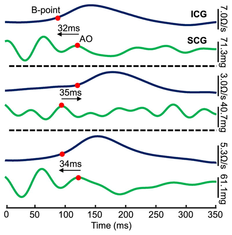
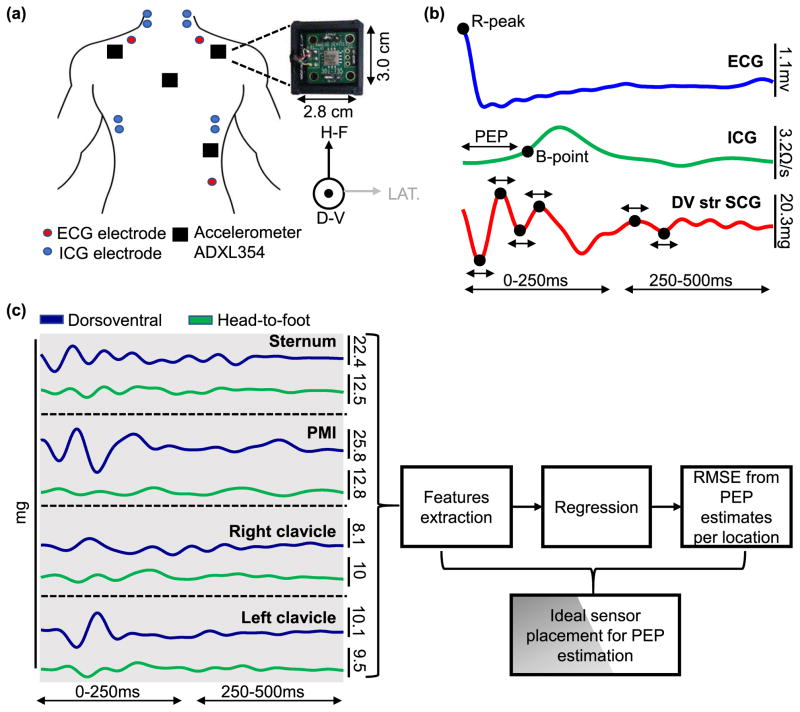
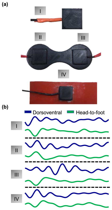
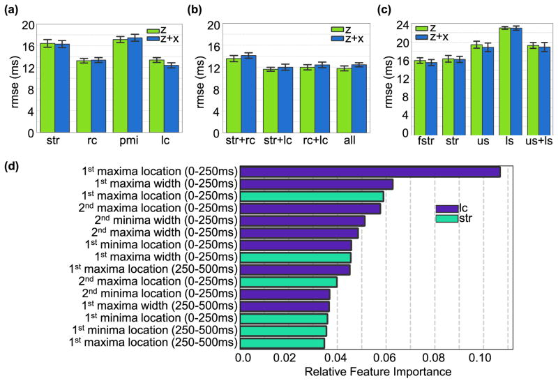
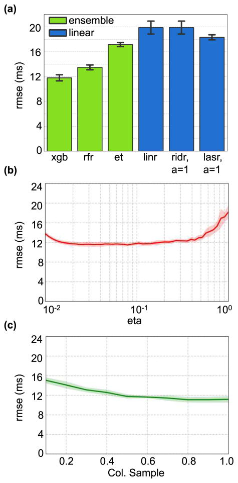
Seismocardiography (SCG), the measurement of local chest vibrations due to the heart and blood movement, is a non-invasive technique to assess cardiac contractility via systolic time intervals such as the pre-ejection period (PEP). Recent studies show that SCG signals measured before and after exercise can effectively classify compensated and decompensated heart failure (HF) patients through PEP estimation. However, the morphology of the SCG signal varies from person to person and sensor placement making it difficult to automatically estimate PEP from SCG and electrocardiogram signals using a global model. In this proof-of-concept study, we address this problem by extracting a set of timing features from SCG signals measured from multiple positions on the upper body. We then test global regression models that combine all the detected features to identify the most accurate model for PEP estimation obtained from the best performing regressor and the best sensor location or combination of locations. Our results show that ensemble regression using XGBoost with a combination of sensors placed on the sternum and below the left clavicle provide the best RMSE = 11.6 ± 0.4 ms across all subjects. We also show that placing the sensor below the left or right clavicle rather than the conventional placement on the sternum results in more accurate PEP estimates.

Keywords: Seismocardiogram; accelerometer; ensemble regression; heart failure; pre-ejection period; sensor fusion; unobtrusive cardiovascular monitoring.


Figures

Fig. 1
Fig. 1
ICG and dorsoventral SCG ensemble averaged traces (n = 5 heartbeats) obtained with the sensor on the sternum for three different subjects. The ICG B-points and SCG AO-points are marked with red circles, and there is a substantial time difference between the two corresponding points for the three subjects: in two cases the ICG B-point occurs first, and in the third case the SCG AO-point occurs first.
Fig. 2
Fig. 2
(a) The experimental setup for Part I of the experiment. Four ADXL354 Accelerometers are placed on the subject, one each at the mid-sternum, below the left and right clavicle, and at the point of maximal impulse. ICG and ECG signals are collected simultaneously. (b) Five beat ensemble averaged traces of ECG, ICG and mid-sternum dorsoventral SCG heartbeats. The ECG R-peak is used as a reference point for beat segmentation, the B-point of the ICG is used to detect aortic valve opening and the R-B interval is used as the ground truth PEP. Peak timing locations and width are extracted from the SCG signal as shown. (c) After extracting the features from the head-to-foot and dorsoventral axes of the SCG signals from all locations, a regression model is used to obtain PEP estimates from the features obtained from a single location, multiple combination of locations, one axis, and both axes. RMSE between the ground truth PEP and every estimate is calculated and the optimal location/ combination of location and axes is determined.
Fig. 3
Fig. 3
(a) Different methods of interfacing the ADXL354 accelerometer with the sternum. (b) SCG signals obtained from each of the different interfacing materials.
Fig. 4
Fig. 4
(a). RMSE from PEP estimated from features obtained from accelerometers placed on the sternum (Str), below the right clavicle (RC), point of maximal impulse (PMI), and below the left clavicle (LC)for both the dorsoventral axis (z-axis) and head-to-foot and dorsoventral axes combined (z+x axes). (b) RMSE from PEP estimated from features obtained from the best performing combination of accelerometer locations. It can be observed that adding more sensors does not substantially reduce the error obtained using one sensor below the left or right clavicle. (c) RMSE from PEP estimated from accelerometers placed on the sternum with different interfacing techniques: in the middle of a silicone rubber sheet placed along on the sternum (fstr), directly on the sternum (Str), and two accelerometers coupled by a rigid plastic mold and placed on the upper sternum (US) and lower sternum (LS). (d) Ranking of best 15 features obtained from the combination of sensors and axis that rendered the lowest RMSE (Str+LC axis z).
Fig. 5
Fig. 5
(a). Comparing RMSE for PEP estimates obtained using ensemble regression models vs. linear regression models on features obtained from SCG signals that performed best with XGBoost (i.e., LC+sternum z-axis). (b) RMSE for PEP estimates obtained using XGBoost on features obtained from LC+sternum z-axis while varying the learning rate parameter (c) RMSE for PEP estimates obtained using XGBoost on features obtained from LC+sternum z-axis while varying the column sample parameter. The shaded regions in parts (b) and (c) indicate the standard deviation since the cross-validation is repeated 50 times.

References

    1. Katz AM. Physiology of the heart. New York: Raven Press; 1977. p. xiii.p. 450.
    1. Go AS, et al. Heart disease and stroke statistics—2014 update: a report from the American Heart Association. Circulation. 2014;129(3):e28. - PMC - PubMed
    1. Huffman MD, et al. Lifetime risk for heart failure among white and black Americans: cardiovascular lifetime risk pooling project. Journal of the American College of Cardiology. 2013;61(14):1510–1517. - PMC - PubMed
    1. Heidenreich PA, et al. Forecasting the future of cardiovascular disease in the United States. Circulation. 2011;123(8):933–944. - PubMed
    1. Inan OT, et al. Using Ballistocardiography to Monitor Left Ventricular Function in Heart Failure Patients. Journal of Cardiac Failure. 2016;22(8):S45.

LinkOut - more resources