Review
. 2018 May 15:12:299.
doi: 10.3389/fnins.2018.00299. eCollection 2018.

Force-Mediating Magnetic Nanoparticles to Engineer Neuronal Cell Function

Affiliations
Review

Force-Mediating Magnetic Nanoparticles to Engineer Neuronal Cell Function

Trevor J Gahl et al. Front Neurosci. .

Abstract

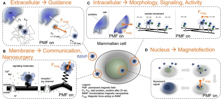
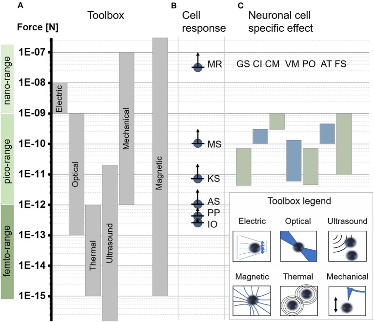
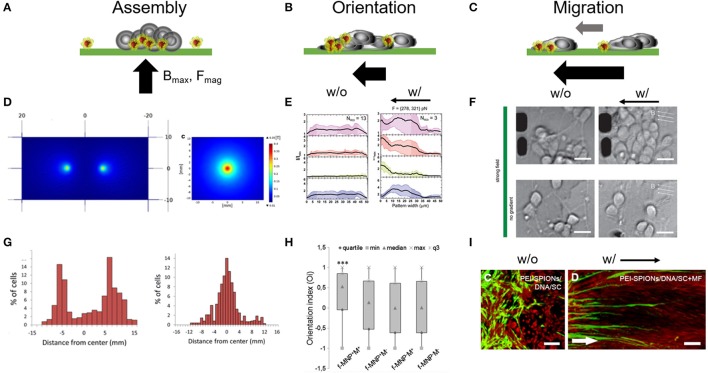
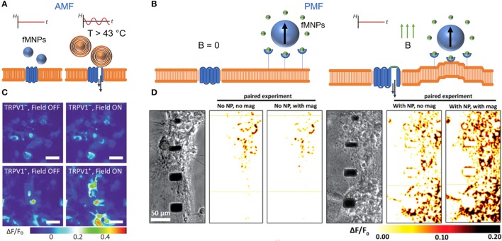
Cellular processes like membrane deformation, cell migration, and transport of organelles are sensitive to mechanical forces. Technically, these cellular processes can be manipulated through operating forces at a spatial precision in the range of nanometers up to a few micrometers through chaperoning force-mediating nanoparticles in electrical, magnetic, or optical field gradients. But which force-mediating tool is more suitable to manipulate cell migration, and which, to manipulate cell signaling? We review here the differences in forces sensation to control and engineer cellular processes inside and outside the cell, with a special focus on neuronal cells. In addition, we discuss technical details and limitations of different force-mediating approaches and highlight recent advancements of nanomagnetics in cell organization, communication, signaling, and intracellular trafficking. Finally, we give suggestions about how force-mediating nanoparticles can be used to our advantage in next-generation neurotherapeutic devices.

Keywords: cell communication; cell guidance; cell polarity; intracellular forces; nanomagnetics; nanoparticles; neurons.


Figures

Figure 1
Figure 1
Force-mediating nanoparticles and their interplay with mammalian cell function. (A) FMNPs as mediators to direct cell guidance through the extracellular space when exposed to a permanent magnetic field gradient (t: time). (B) FMNPs associated to the cell membrane can control receptor functionality, stimulate cell communication, or perform local cell surgeries. (C) Positioning of fMNPs inside cells can establish protein gradients and modulate vesicle dynamics. (D) Localizing FMNPs to the cell nucleus is utilized to genetically modify cells, here demonstrated through the translation of a fluorescent protein (e.g., GFP, or eGFP).
Figure 2
Figure 2
Force scales relevant to subcellular applications. (A) Force-generating toolbox to manipulate subcellular forces. Force direction and amplitude are controlled through physical concepts based on electric field gradients = electric, optical tweezers = optical, acoustic tweezers = ultrasound, magnetic tweezers = magnetic, thermal tweezers = thermal, or mechanical actuation = mechanical. (Jeney et al., ; Neuman and Nagy, ; Mosconi et al., 2011). (B) Minimum reported force values required for different cell effects based on computational or experimental models including membrane rupture (MR), (Almeida and Vaz, 1995) microtubules stretching (MS), (Dogterom and Yurke, ; Brangwynne, 2006) kinesin motor stalling (KS), (Svoboda and Block, ; Visscher et al., 1999) actin stalling (AS), (Footer et al., 2007) protein polymerization (PP), (Footer et al., 2007) ion channel opening and thermal fluctuation (IO). (Meister, 2016) (C) Overview of experimentally reported force ranges indicating significant changes in neuronal cell function and morphology induced through a force stimulus. The list highlights general reported mechanical sensitivity for neurons (GS), (Zablotskii et al., 2016a,b) force mediated calcium induction (CI), (Calabrese et al., ; Matthews et al., ; Maneshi et al., ; Tay et al., 2016a) force mediated cell migration/displacement (CM), (Kunze et al., 2015) force mediated modulation of vesicle motion (VM), (Kunze et al., 2017) force mediated protein positioning (PO), (Kunze et al., 2015) force mediated axon towing and stretching (AT), (Bray, ; Suter and Miller, 2011) and force mediated filopodia/growth cone stretching (FS). (Franze, 2013).
Figure 3
Figure 3
Magnetic forces as cell patterning mediators. Tuning magnetic force amplitudes (Fmag), the position of the maximal magnetic field (Bmax) and the orientation of the magnetic field pole indicated through the black arrow provides a versatile approach for cell assembly. (A–C) Schematic represents different magnetic field gradient orientations and magnitudes and its impact on cell assembly and organization. (D) Single vs. two pole magnetic field gradient spots for positioning of cells. Reproduced with permission from Marcus et al. (2016), Copyright © 2016, BioMed Central. (E) Fluorescence distribution plots taken from primary cortical neuron cultures show a shift of intracellular markers toward left oriented magnetic gradient forces. w/o, no magnetic field; w/, with magnetic field. Reproduced with permission from Kunze et al. (2015). (F) Primary cortical neurons dissociated from rat brain tissues (E18) were cultured on poly-l-lysine surfaces and exposed to fMNPs after being 24 h in culture. These neurons grow and form neurite networks under magnetic fields and start migration toward magnetic field poles under strong magnetic forces (> 250 pN). Scale bar = 12 μm. Reproduced with permission from Kunze et al. (2015), Copyright © 2015, American Chemical Society. (G) Histogram plots of accumulated neuron-like cells which were cultured above the single and two pole patterns, respectively. (H) Orientation index extracted from PC12 that were observed to align in parallel to magnetic field orientation after being cultured with fMNPs. f-MNP-M+, fMNPs with magnetic field; f-MNP-M, fMNPs without magnetic field. Reproduced with permission from Riggio et al. (2014), Copyright © 2014, Elsevier Inc. (I) Schwann cells migrate into astrocyte-rich region under an oriented magnetic field gradient after internalizing PEI-fMNPs (PEI-SPIONs). White arrow indicates direction of magnetic pole. Scale bar = 100 μm. Reproduced with permission from Xia et al. (2016), Copyright © 2016, Dove Medical Press Limited.
Figure 4
Figure 4
Controlling calcium influx with alternating (AMF) and permanent magnetic fields (PMF). (A) Heat stimulation of TRPV1 through fMNP-mediated heat induction in AMF. (B) FMNP-mediated calcium channel activation via membrane bending in PMF. “B = 0” indicates no magnetic field. (C) False color heat maps show changes in fluorescently-labeled intracellular calcium concentration in TRPV1- and TRPV+ HEK293FT cells before and during AMF stimulation. Scale bar = 50 μm. Reproduce with permission from Chen et al. (2015), Copyright © 2015, American Association for the Advancement of Science. (D) False color heat maps show changes in fluorescently-labeled intracellular calcium influx in primary cortical neurons (E18, rat) with and without fMNPs and with and without PMF stimulation. Reproduce with permission from Tay et al. (2016a), Copyright © 2016, American Chemical Society.
Figure 5
Figure 5
Force-mediated protein sorting inside cells across different levels of complexity in cell morphology. (A–C) Schematic representation of different levels of cell complexity ranging from almost perfectly round to highly branched structures. (D) Microtubules nucleation in artificial, micro-scaled lipid droplets de-centralize the nucleation zone through the application of magnetic field gradient. Magnetic forces off-center nucleation position of microtubules through repulsion. Forces were estimated in the femtonewton range. (E) Altered protein positioning (HaloTag-eGFP) through nanomagnetic forces operated by magnetic tweezers in HeLa cells. Scale bar = 10 μm. HTL-sMNPs, HaloTag-ligand-silica-based magnetic nanoparticles. (F) Primary cortical neurons with superparamagnetic nanoparticles re-assemble Tau proteins toward the magnetic field gradient when exposed to a permanent magnetic field. Scale bar = 16 μm. (G) Microtubules nucleation position of RanGTP-magnetic nanoparticles (Ran-NPs) without (Off) and with (On) magnetic forces. (D,G) Reproduced with permission from Bonnemay et al. (2013), Copyright © 2013, American Chemical Society. (H) Surface intensity plot shows correlation between nanoparticles (HTL-sMNPs = sMNPs) and protein assembly (HT-eGFP) in transfected HeLa cells dropping away from the magnetic tip. (E,H) Reproduced with permission from Etoc et al. (2015), Copyright © 2015, American Chemical Society. (I) Histogram plot for the nanomagnetic force range were protein assembly was significant different from its native distribution. (F,I) Adapted from Kunze et al. (2015), Copyright © 2015, American Chemical Society. (J) Force-mediated local activation actin cytoskeleton dynamics through dragging Rho-family GTPases proteins. Reproduced with permission from Levskaya et al. (2009), Copyright © 2009, Springer Nature.
Figure 6
Figure 6
Dynamic behavior of intracellular vesicles is sensitive to force-mediated changes inside and outside of neurons. Changes in vesicle dynamics are studied through (A) a stretchable or (B) a localized nanomagnetic cell culture platform. (C) The stretchable cell culture surface imposes a uniform elongation on adherent parts of the cell body and cytoskeleton. (D) Histogram shows differences in force-mediated active vs. axonal forward (anterograde) transport. (E) On chip method to mechanically interfere with vesicle dynamics inside neurons through nanomagnetic forces. (F) Estimated nanomagnetic force map. (G) Stretch mediates the increase of synaptic vesicles in neuromuscular synapses in Drosophila embryonic motor neurons. (H) Nanoparticle-laden lipid vesicles in primary cortical neurons alter their movement pattern under magnetic forces. (I) Extracted vesicle tracks without (no M) and with (w M) nanomagnetic forces. (C,D,G) Reproduced with permission from Ahmed et al. (2012,2013), Copyright © 2012, Royal Society of Chemistry. (E,F,H,I) were adapted and reproduced with permission from Kunze et al. (2017), Copyright © 2017, Royal Society of Chemistry.
Figure 7
Figure 7
Suggested future studies should address fundamental aspects of how nanomagnetic forces associate with cellular structures or how nanomagnetic force stimulation can be integrated into therapeutic and translational approaches. (A) Protein-protein interactions are suggested to play a dominant role in nanomagnetic force activation and may determine how much force is required and how sensitive cells are to a biomechanical stimulus at the membrane. Depending on the surface functionality nanoparticles may interact with the cellular membrane in a weak associative or on a strong bound connection. The strong bound connection suggests an immediate deformation of the membrane resulting in a short lag time to trigger a specific intracellular downstream process after a stimulus occurred. In contrast to the strong connection, the weaker associative connection may lead to a longer lag time or result in no further activation of downstream processes. (B) Other research efforts should focus on integrating nanomagnetic force stimulation into current neuromodulation tools, tissue engineering, organ functionality and translation into diagnostics, patient-specific therapeutics, or treatment predictions.

Similar articles

Cited by

References

    1. Adams C. F., Pickard M. R., Chari D. M. (2013). Magnetic nanoparticle mediated transfection of neural stem cell suspension cultures is enhanced by applied oscillating magnetic fields. Nanomedicine 9, 737–741. 10.1016/j.nano.2013.05.014 - DOI - PubMed
    1. Ahmed W. W., Kural M. H., Saif T. A. (2010). A novel platform for in situ investigation of cells and tissues under mechanical strain. Acta Biomater. 6, 2979–2990. 10.1016/j.actbio.2010.02.035 - DOI - PMC - PubMed
    1. Ahmed W. W., Li T. C., Rubakhin S. S., Chiba A., Sweedler J. V., Saif T. A. (2012). Mechanical tension modulates local and global vesicle dynamics in neurons. Cell. Mol. Bioeng. 5, 155–164. 10.1007/s12195-012-0223-1 - DOI - PMC - PubMed
    1. Ahmed W. W., Williams B. J., Silver A. M., Saif T. A. (2013). Measuring nonequilibrium vesicle dynamics in 804 neurons under tension. Lab. Chip 13, 570-578. 10.1039/c2lc41109a - DOI - PubMed
    1. Allen Liu P. (2016). Biophysical tools for cellular and subcellular mechanical actuation of cell signaling Biophys. J. 111, 1112–1118. 10.1016/j.bpj.2016.02.043 - DOI - PMC - PubMed

LinkOut - more resources