. 2018 May 15:9:437.
doi: 10.3389/fphar.2018.00437. eCollection 2018.

Bushen Huoxue Recipe Alleviates Implantation Loss in Mice by Enhancing Estrogen-Progesterone Signals and Promoting Decidual Angiogenesis Through FGF2 During Early Pregnancy

Affiliations

Bushen Huoxue Recipe Alleviates Implantation Loss in Mice by Enhancing Estrogen-Progesterone Signals and Promoting Decidual Angiogenesis Through FGF2 During Early Pregnancy

Jiahui Ding et al. Front Pharmacol. .

Abstract

Bushen Huoxue recipe (BSHXR) is a classic Chinese herbal prescription for nourishing the kidney and activating blood circulation. It consists of six herbs: Astragali radix, Angelicae sinensis radix, Ligustici Chuanxiong Rhizoma, Cuscutae semen, Taxilli Herba, and Dipsaci Radix, and the main active constituents of BSHXR are ferulic acid, calycosin-7-glucopyranoside, hyperoside, quercitrin, and asperosaponin VI. In clinical practice, BSHXR is traditionally used to treat failed pregnancy and its complications. However, little is known about the underlying mechanism of BSHXR for the treatment of implantation loss during early pregnancy. In the current study, controlled ovarian hyperstimulation was induced in mice as our implantation loss model, and we evaluated the effects of BSHXR on implantation, decidualization, decidual angiogenesis, and reproductive outcome. We showed that BSHXR could regulate the supraphysiological levels of serum estrogen and progesterone observed in these mice, and also act on estrogen and progesterone receptors in the stroma and epithelium. BSHXR also enhanced FGF2 expression in the vascular sinus folding area of the decidua, thus potentially reducing implantation loss during early pregnancy and contributing to placentation and survival of the fetuses. Taken together, our findings provide scientific evidence for the application of BSHXR in the clinic as a treatment for implantation loss during early pregnancy, and warrant further investigation of BSHXR as an effective treatment for failed pregnancy and its complications.

Keywords: Bushen Huoxue recipe; FGF2; decidual angiogenesis; early pregnancy loss; estrogen; implantation loss; progesterone; traditional Chinese medicine.


Figures

FIGURE 1
FIGURE 1
The graphic illustration of experiment. E, embryo; ER, estrogen receptor; PR, progesterone receptor.
FIGURE 2
FIGURE 2
The HPLC fingerprints of reference standards (A), BSHXR (B), and BSHXR from three batches (C). Peak number and identity: 1, ferulic acid; 2, calycosin-7-Glucopyranoside; 3, hyperoside; 4, quercitrin; 5, asperosaponin VI.
FIGURE 3
FIGURE 3
Visible external morphology of uterus showing implantations on D6 (A) and on D8 (B). Arrows indicate implantation sites.
FIGURE 4
FIGURE 4
Effect of BSHXR on the morphology of ovary and serum hormones. (A) Morphology analysis of ovary section (HE, ×40), lines indicate maximum diameters of corpus luteum. Bars = 500 μm. (B) Measurement of bilateral ovary weight on D5, D6, and D8, N = 9–10. (C) Serum estradiol-17β levels on D5, D6, and D8, N = 8–10. (D) Serum progesterone levels on D5, D6, and D8, N = 8–10. Data represent as mean ± SD. P < 0.05, ∗∗P < 0.01, and ∗∗∗P < 0.001 when compared to control group. #P < 0.05, ##P < 0.01, and ###P < 0.001 when compared to model group.
FIGURE 5
FIGURE 5
Morphology analysis of uterine section on D5, D6, and D8. The right pictures are the higher magnification images of the black boxes or ellipses on the left. (A) The development of glands on D5. Arrows indicate glands. (B) Formation of PDZ on D6. Arrows indicate decidual cells. (C) Formation of SDZ and surrounding blood vessel of embryo on D8. Arrows indicate blood vessels and arrowheads indicate decidual cells. E, embryo; L, lumen; G, gland; PDZ, primary decidual zone; SDZ, secondary decidual zone; M, mesometrial; AM, anti-mesometrial; VSF, vascular sinus folding. Original magnifications and scale bar were shown in images.
FIGURE 6
FIGURE 6
ERα expression in the uterus on D5, D6 and D8. (A) Immunohistochemistry of ERα in paraffin sections of the uterus on D5 (A), D6 (B), and D8 (C). For each group (control, model, BSHXR1, and BSHXR2), a low magnification image of the uterus is depicted, as well as higher magnification images of the lumen (L), gland (G), and stroma (S). E, embryo; M, mesometrial; AM, anti-mesometrial; L, lumen; G, gland; S, stroma. Arrows indicate glands. Original magnification and scale bar were shown in images. (D) Protein levels of ERα in pregnant uterus (remove embryo) by western blot analysis (n = 3). (E) Quantification of ERα protein expression. (F) mRNA levels of ERα in pregnant uterus (remove embryo) by qRT-PCR (n = 5). Data represent mean ± SD. β-actin was used as the reference. P < 0.05, ∗∗P < 0.01, and ∗∗∗P < 0.001 when compared to control group. #P < 0.05, ##P < 0.01, and ###P < 0.001 when compared to model group.
FIGURE 7
FIGURE 7
PR expression in the uterus on D5, D6 and D8. (A) Immunohistochemistry of PR in paraffin sections of the uterus on D5 (A), D6 (B), and D8 (C). For each group (control, model, BSHXR1, and BSHXR2), a low magnification image of the uterus is depicted, as well as higher magnification images of the lumen (L), gland (G) and stroma (S). E, embryo; M, mesometrial; AM, anti-mesometrial; L, lumen; G, gland; S, stroma. Arrows indicate glands. Original magnification and scale bar were shown in images. (D) Protein levels of PR in pregnant uterus (remove embryo) by western blot analysis (n = 3). (E) Quantification of PR protein expression. (F) mRNA levels of PR in pregnant uterus (remove embryo) by qRT-PCR (n = 5). Data represent mean ± SD. β-actin was used as the reference. ∗∗P < 0.01 when compared to control group. #P < 0.05 and ##P < 0.01 when compared to model group.
FIGURE 8
FIGURE 8
Decidual angiogenesis of the pregnant uterus on D5, D6, and D8. Immunohistochemistry staining with cluster of differentiation 31 (CD31). The pictures on the right are the higher magnification images of VSF. Arrow represents the variable-sized blood vessels. E, embryo; L, lumen; M, mesometrial; AM, anti-mesometrial; VSF, vascular sinus folding. Original magnification and scale bar were shown in the pictures.
FIGURE 9
FIGURE 9
VEGFA expression in the uterus on D5, D6, and D8. (A) Immunohistochemistry of VEGFA in paraffin sections of the uterus. The right pictures are the higher magnification images of the black boxes on the left. Arrow indicates the positive staining. L, lumen; G, gland; M, mesometrial; AM, anti-mesometrial; VSF, vascular sinus folding. Original magnification and scale bar were shown in the pictures. (B) Protein levels of VEGFA in pregnant uterus (remove embryo) by western blot analysis (n = 3). (C) Quantification of VEGFA protein expression. (D) mRNA levels of VEGFA in pregnant uterus (remove embryo) by qRT-PCR (n = 5). Data represent mean ± SD. β-actin was used as the reference.
FIGURE 10
FIGURE 10
ANGPT2 expression in the uterus on D5, D6, and D8. (A) Immunohistochemistry of ANGPT2 in paraffin sections of the uterus. The right pictures are the higher magnification images of the black boxes on the left. Arrow indicates the positive staining. L, lumen; E, embryo; M, mesometrial; AM, anti-mesometrial; VSF, vascular sinus folding. Original magnification and scale bar were shown in the pictures. (B) Protein levels of ANGPT2 in pregnant uterus (remove embryo) by western blot analysis (n = 3). (C) Quantification of ANGPT2 protein expression. (D) mRNA levels of ANGPT2 in pregnant uterus (remove embryo) by qRT-PCR (n = 5). Data represent mean ± SD. β-actin was used as the reference.
FIGURE 11
FIGURE 11
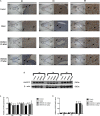
FGF2 expression in the uterus on D5, D6, and D8. (A) Immunohistochemistry of FGF2 in paraffin sections of the uterus. The right pictures are the higher magnification images of the black boxes on the left. Arrow indicates the positive staining. L, lumen; E, embryo; M, mesometrial; AM, anti-mesometrial; VSF, vascular sinus folding. Original magnification and scale bar were shown in the pictures. (B) Protein levels of FGF2 in pregnant uterus (remove embryo) by western blot analysis (n = 3). (C) Quantification of FGF2 protein expression. (D) mRNA levels of FGF2 in pregnant uterus (remove embryo) by qRT-PCR (n = 5). Data represent mean ± SD. β-actin was used as the reference. ∗∗P < 0.01 and ∗∗∗P < 0.001 when compared to control group. #P < 0.05, ##P < 0.01, and ###P < 0.001 when compared to model group.

References

    1. Adams N. R., DeMayo F. J. (2015). The role of steroid hormone receptors in the establishment of pregnancy in rodents. 216 27–49. 10.1007/978-3-319-15856-3_3 - DOI - PubMed
    1. Bindali B. B., Kaliwal B. B. (2002). Anti-implantation effect of a carbamate fungicide mancozeb in albino mice. 40 191–197. 10.2486/indhealth.40.191 - DOI - PubMed
    1. Carson D. D., Bagchi I., Dey S. K., Enders A. C., Fazleabas A. T., Lessey B. A., et al. (2000). Embryo implantation. 223 217–237. 10.1006/dbio.2000.9767 - DOI - PubMed
    1. Cha J., Sun X., Dey S. K. (2012). Mechanisms of implantation: strategies for successful pregnancy. 18 1754–1767. 10.1038/nm.3012 - DOI - PMC - PubMed
    1. Demir R., Yaba A., Huppertz B. (2010). Vasculogenesis and angiogenesis in the endometrium during menstrual cycle and implantation. 112 203–214. 10.1016/j.acthis.2009.04.004 - DOI - PubMed

LinkOut - more resources