. 2018 May 18:9:517.
doi: 10.3389/fphar.2018.00517. eCollection 2018.

Coptisine Induces Apoptosis in Human Hepatoma Cells Through Activating 67-kDa Laminin Receptor/cGMP Signaling

Affiliations

Coptisine Induces Apoptosis in Human Hepatoma Cells Through Activating 67-kDa Laminin Receptor/cGMP Signaling

Li Zhou et al. Front Pharmacol. .

Abstract

Hepatocellular carcinoma (HCC) is the most common primary cancer of the liver. Hence, new anti-liver cancer treatment strategies need to be urgently developed. Coptisine is a natural alkaloid extracted from rhizoma coptidis which exhibits anticancer activity in various preclinical models, including liver cancer. However, the molecular mechanisms underlying the anti-liver cancer effects of coptisine remains unclear. We used flow cytometry to assess the binding of coptisine to 67LR expressed on the surface of SMMC7721, HepG2, LO2 and H9 cells. Then SMMC7721, HepG2 and BEL7402 cells, belonging to the HCC cell lines, were treated with coptisine. The cell viability was detected using a cell counting kit-8 assay. Apoptosis was evaluated using flow cytometry and transferase-mediated dUTP nick-end labeling (TUNEL) assay. Apoptotic-related proteins and tumor death receptor 67-kDa laminin receptor (67LR) were detected using Western blot analysis. The cyclic guanosine 3',5'-monophosphate (cGMP) concentration was determined using enzyme-linked immunosorbent assay. sh67LR lentivirus, anti67LR antibody, and cGMP inhibitor NS2028 were used to determine how a 67LR/cGMP signaling pathway regulated coptisine-induced apoptosis. Tumor growth inhibited by coptisine was confirmed in a SMMC7721 cell xenograft mouse model. Coptisine selectively exhibited cell viability in human hepatoma cells but not in normal human hepatocyte cell line LO2 cells. Coptisine promoted SMMC7721 and HepG2 cell apoptosis by increasing 67LR activity. Both 67LR antibody and sh67LR treatment blocked coptisine-induced apoptosis and inhibition of cell viability. Coptisine upregulated the expression of cGMP. Moreover, cGMP inhibitor NS2028 significantly decreased coptisine-induced apoptosis and inhibition of cell viability. In vivo experiments confirmed that coptisine could significantly suppress the tumor growth and induce apoptosis in SMMC7721 xenografts through a 67LR/cGMP pathway. Coptisine-mediated 67LR activation may be a new therapeutic strategy for treating hepatic malignancy.

Keywords: 67LR; apoptosis; cGMP; coptisine; hepatocellular carcinoma.


Figures
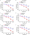
FIGURE 1
FIGURE 1
Coptisine inhibited cell viability in SMMC7721, HepG2 and BEL7402 cells but not in LO2 cells. (A) SMMC7721 cells, (C) HepG2 cells, (E) BEL7402 cells and LO2 cells were treated with indicated concentrations of coptisine for 24 h, and cell viability was detected using CCK-8 assay. P < 0.05 and ∗∗P < 0.01 compared with the untreated group (control) of the same cells. #P < 0.05 compared with the same operated group of SMMC7721 cells. (B) SMMC7721 cells, (D) HepG2 cells, (F) BEL7402 cells and LO2 cells were treated with 50 μM coptisine for 0–24 h, and cell viability was detected using CCK-8 assay. P < 0.05 and ∗∗P < 0.01 compared with the 0-h group of the same cells. #P < 0.05 compared with the same group of SMMC7721 cells. The results were representative of three independent experiments. Error bars represent mean ± SD.
FIGURE 2
FIGURE 2
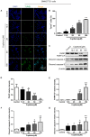
Coptisine concentration-dependent induced apoptosis in SMMC7721 cells. (A) The cells were treated with indicated concentrations of coptisine for 24 h. The morphologic changes in apoptotic cells were evaluated using TUNEL staining. Scale bar represents 150 μm. (B) The percentages of TUNEL-positive cells were calculated using ImageJ software. (C) Total protein lysates were collected after coptisine treatment, and the expression of proteins was detected using Western blotting analysis with the indicated antibodies. PARP-CF means the C-terminal catalytic fragment of PARP. (D–G) Relative densities of proteins were analyzed using ImageJ software. The results were representative of three independent experiments. Error bars represent the mean ± SD. P < 0.05 and ∗∗P < 0.01 compared with control.
FIGURE 3
FIGURE 3
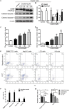
Coptisine concentration-dependent induced apoptosis in HepG2 cells and Coptisine could bind to the surface of hepatoma carcinoma cells. (A) After HepG2 cells were treated with indicated concentrations of coptisine for 24 h, total protein lysates were collected, and the expression of proteins was detected using Western blotting analysis with the indicated antibodies. PARP-CF means the C-terminal catalytic fragment of PARP. (B–D) Relative densities of proteins were analyzed using ImageJ software. The results were representative of three independent experiments. Error bars represent the mean ± SD. P < 0.05 and ∗∗P < 0.01 compared with control. (E) 50 mM Coptisine was labeled by 0.5 mM FITC for 30 min (FITC-labeled coptisine), and then added to SMMC7721 cells, HepG2 cells, LO2 cells and H9 cells respectively. 1 μg/ml PI was used to determine living cells. FITC-labeled coptisine was heat-inactivated as a negative control. Cells were filtrated by flow cytometry. (F) The percentages of FITC positive cells were counted using FlowJo software (n = 3). (G) The percentages of PI positive cells were counted (n = 3). P < 0.05 and ∗∗P < 0.01 compared with the appointed group.
FIGURE 4
FIGURE 4
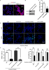
Coptisine promoted SMMC7721 cell apoptosis by increasing 67LR activity. (A) LO2 cells and SMMC7721 cells were separately collected and stained with anti-67LR (red) and DAPI (blue) to identify the expression of 67LR using immunofluorescence. The scale bar represents 50 μm. (B,C) The expression of 67LR protein was tested in scramble shRNA SMMC7721 and sh67LR SMMC7721 cells using Western blot analysis. The relative density of 67LR expression was analyzed using ImageJ software. P < 0.05 compared with the scramble shRNA group. (D,E) Anti-67LR and sh67LR models were used to detect cell apoptosis after coptisine (50 μM) treatment. Then, morphologic changes in apoptotic cells were evaluated using TUNEL staining. Scale bar represents 150 μm. The percentages of TUNEL-positive cells were calculated using ImageJ software. ∗∗P < 0.01. (F) Cell viability was tested using CCK-8 assay. ∗∗P < 0.01; NS means no statistical significance. The results were representative of three independent experiments. All data are represented as mean ± SD.
FIGURE 5
FIGURE 5
67LR activity significantly influenced the effect of coptisine on SMMC7721 cell apoptosis. (A,B) Apoptotic cells were quantified using flow cytometry after staining with Annexin V–APC and DAPI according to the indicated groups. Also, the percentages of apoptotic cells were counted. ∗∗P < 0.01 compared with the appointed group. Values represent mean ± SD with three replicates.
FIGURE 6
FIGURE 6
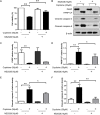
Coptisine promoted HepG2 cell apoptosis by increasing 67LR activity. (A) Anti-67LR and sh67LR models were used to detect cell apoptosis after coptisine (50 μM) treatment. Then, morphologic changes in apoptotic cells were evaluated using TUNEL staining. The percentages of TUNEL-positive cells were calculated using ImageJ software. ∗∗P < 0.01. (B) Total protein lysates were collected after treatment, then the expression of proteins was tested by using Western blotting analysis with the indicated antibodies. PARP-CF means the C-terminal catalytic fragment of PARP. (C–F) Relative densities of proteins were analyzed using ImageJ software. The results were representative of three independent experiments. Error bars represent the mean ± SD. P < 0.05 and ∗∗P < 0.01 compared with the appointed group.
FIGURE 7
FIGURE 7
Inhibiting the expression of cGMP could obviously reduce coptisine-induced apoptosis. (A) The viability of SMMC7721 cells was detected using CCK-8 assay according to the indicated groups. NS2028 (5 μM) was used to inhibit the expression of cGMP. (B) Total protein lysates were collected after coptisine and (or) NS2028 treatment, and then the expression of proteins was detected using Western blot analysis using the indicated antibodies. PARP-CF means the C-terminal catalytic fragment of PARP. (C–F) The relative densities of these proteins were analyzed using ImageJ software. P < 0.05 and ∗∗P < 0.01 compared with the appointed group (n = 3 per group for all the studies). All data are expressed as mean ± SD.
FIGURE 8
FIGURE 8
Coptisine induced cGMP upregulation after activating 67LR in SMMC7721 cells. (A,B) Apoptotic cells were quantified using flow cytometry after staining with Annexin V–APC and DAPI according to the indicated groups. Also, the percentages of apoptotic cells were counted (n = 3). (C) The concentration of induced cGMP was measured using an ELISA kit after coptisine and (or) NS2028 treatment (n = 6). P < 0.05 and ∗∗P < 0.01 compared with the appointed group. All data are expressed as mean ± SD.
FIGURE 9
FIGURE 9
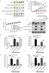
Coptisine inhibited tumor growth and induced apoptosis in a SMMC7721 xenograft animal model. A total of 50 nude mice were inoculated with SMMC7721 cells and then randomly divided into 5 groups (n = 10). (A) Images were exhibited for five representative tumors from each group after 35 days of treatment. (B) Tumor volumes were measured according to the indicated intervals. Data are expressed as mean ± SD. P < 0.05 and ∗∗P < 0.01 compared with the same-day results of the control group. #P < 0.05 compared with the same-day results of the coptisine + IgG group. @P < 0.05 and @@P < 0.01 compared with the same-day results of the coptisine + scramble shRNA group. (C) Changes in the body weight of mice were recorded during the 35 days of treatment. (D–H) Tumors from all the five groups were lysed and collected to detect the expression of objective proteins using Western blot analysis. PARP-CF means the C-terminal catalytic fragment of PARP. The relative densities of these proteins were analyzed using ImageJ software. P < 0.05, ∗∗P < 0.01, and #P < 0.05 (n = 3 per group for all the studies. All data are expressed as mean ± SD.
FIGURE 10
FIGURE 10
Coptisine induced apoptosis through a 67LR-dependent pathway in a SMMC7721 xenograft animal model. (A) Tumors from the coptisine + scramble shRNA and coptisine + sh67LR groups were fixed and stained with H&E to inspect tumor cell morphology. Also, immunohistochemical analysis was used to evaluate the levels of apoptotic -related proteins. Scale bar represents 200 μm. (B) An illustration of the molecular mechanism of coptisine-induced apoptosis. Coptisine induced 67LR functional activation and promoted sGC conversion to generate cGMP, resulting in cGMP upregulation, then caspase activation, and finally apoptosis.

References

    1. Boege Y., Malehmir M., Healy M. E., Bettermann K., Lorentzen A., Vucur M. (2017). A dual role of caspase-8 in triggering and sensing proliferation-associated DNA damage, a key determinant of liver cancer development. 32 342.e10–359.e10. 10.1016/j.ccell.2017.08.010 - DOI - PMC - PubMed
    1. Dorchies O. M., Wagner S., Buetler T. M., Ruegg U. T. (2009). Protection of dystrophic muscle cells with polyphenols from green tea correlates with improved glutathione balance and increased expression of 67LR, a receptor for (-)-epigallocatechin gallate. 35 279–294. 10.1002/biof.34 - DOI - PubMed
    1. Fan L., Xiao Q., Chen Y., Chen G., Duan J., Tao W. (2017). Pekinenin E inhibits the growth of hepatocellular carcinoma by promoting endoplasmic reticulum stress mediated cell death. 8:424. 10.3389/fphar.2017.00424 - DOI - PMC - PubMed
    1. Gravitz L. (2014). Liver cancer. 4:516. 10.1038/516S1a - DOI - PubMed
    1. Hu Y. R., Ma H., Zou Z. Y., He K., Xiao Y. B., Wang Y., et al. (2017). Activation of Akt and JNK/Nrf2/NQO1 pathway contributes to the protective effect of coptisine against AAPH-induced oxidative stress. 85 313–322. 10.1016/j.biopha.2016.11.031 - DOI - PubMed

LinkOut - more resources