. 2018 May 9:9:513.
doi: 10.3389/fphys.2018.00513. eCollection 2018.

Validated Computational Model to Compute Re-apposition Pressures for Treating Type-B Aortic Dissections

Affiliations

Validated Computational Model to Compute Re-apposition Pressures for Treating Type-B Aortic Dissections

Aashish Ahuja et al. Front Physiol. .

Abstract

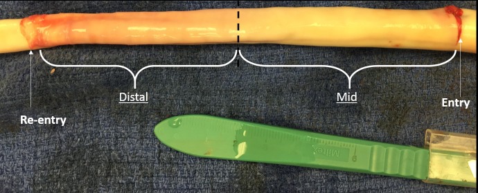
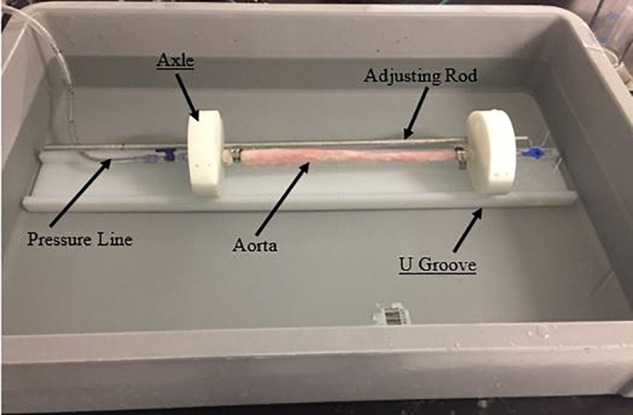
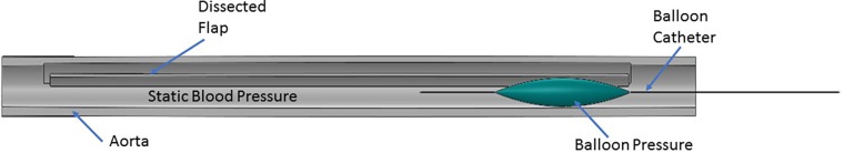
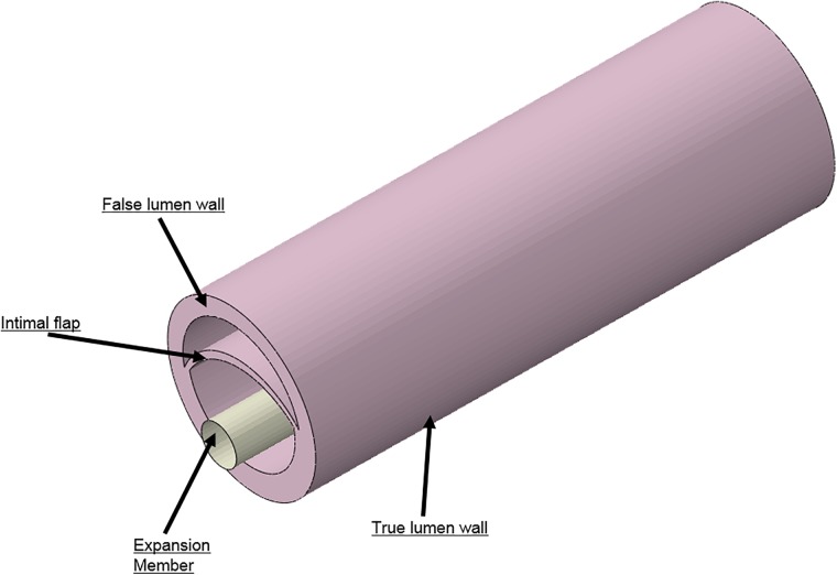
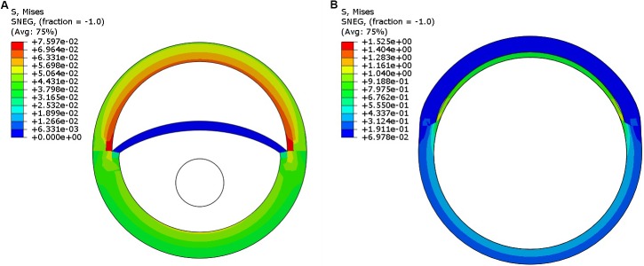
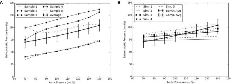
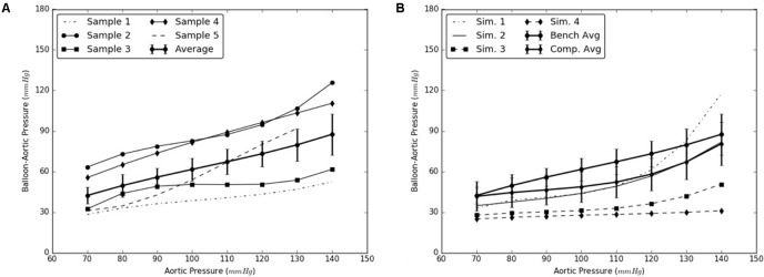
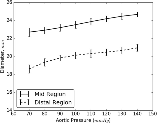
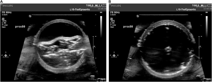
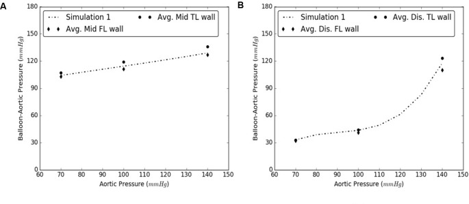
The use of endovascular treatment in the thoracic aorta has revolutionized the clinical approach for treating Stanford type B aortic dissection. The endograft procedure is a minimally invasive alternative to traditional surgery for the management of complicated type-B patients. The endograft is first deployed to exclude the proximal entry tear to redirect blood flow toward the true lumen and then a stent graft is used to push the intimal flap against the false lumen (FL) wall such that the aorta is reconstituted by sealing the FL. Although endovascular treatment has reduced the mortality rate in patients compared to those undergoing surgical repair, more than 30% of patients who were initially successfully treated require a new endovascular or surgical intervention in the aortic segments distal to the endograft. One reason for failure of the repair is persistent FL perfusion from distal entry tears. This creates a patent FL channel which can be associated with FL growth. Thus, it is necessary to develop stents that can promote full re-apposition of the flap leading to complete closure of the FL. In the current study, we determine the radial pressures required to re-appose the mid and distal ends of a dissected porcine thoracic aorta using a balloon catheter under static inflation pressure. The same analysis is simulated using finite element analysis (FEA) models by incorporating the hyperelastic properties of porcine aortic tissues. It is shown that the FEA models capture the change in the radial pressures required to re-appose the intimal flap as a function of pressure. The predictions from the simulation models match closely the results from the bench experiments. The use of validated computational models can support development of better stents by calculating the proper radial pressures required for complete re-apposition of the intimal flap.

Keywords: aortic dissection; bench tests; finite element analysis; porcine aorta; re-apposition pressure; simulation models.


Figures

FIGURE 1
FIGURE 1
An inverted aorta with an imposed dissection. An entry was initially created in the descending thoracic aorta and propagated using forceps to the distal region of the aorta where a pocket of re-entry was created. Tissue specimens from two regions (mid and distal) were extracted and tested on planar biaxial testing machine for material characterization.
FIGURE 2
FIGURE 2
Fixture and setup for conducting bench tests on dissected aorta.
FIGURE 3
FIGURE 3
Representation of the protocol followed during the bench test. A balloon catheter was inserted into the true lumen (TL). The balloon was inflated with water which applied bending forces to either the mid or distal region of the flap in the presence of static aortic pressure. The balloon pressure on re-apposition of flap was recorded using a pressure transducer.
FIGURE 4
FIGURE 4
Different geometries required to simulate aortic dissection are developed as 3D computer aided design (CAD) models and imported into finite element analysis (FEA) software.
FIGURE 5
FIGURE 5
(A) Pressurization of the dissected aorta to an internal pressure of 100 mmHg. (B) Expansion member was distended to achieve full re-apposition of the intimal flap.
FIGURE 6
FIGURE 6
(A) Bench test measurements for re-apposition of mid flap against the mid false lumen (FL) wall. (B) Computational measurements for re-apposition of mid flap and its comparison with bench results.
FIGURE 7
FIGURE 7
(A) Bench test measurements for re-apposition of distal flap against the distal false lumen (FL) wall. (B) Computational measurements for re-apposition of distal flap and its comparison with bench results.
FIGURE 8
FIGURE 8
Resulting diameter of the mid and distal region of dissected aorta on complete re-apposition of intimal flap.
FIGURE 9
FIGURE 9
Ultrasound images capturing various stages: (A) crimped and (B) expanded, of a CODA balloon under aortic pressure of 80 mmHg.
FIGURE 10
FIGURE 10
Comparison between results from Sample #1 and averages of samples #2-5 for (A) Mid, and (B) Distal dissections.

Similar articles

Cited by

References

    1. Alves C. M. R., da Fonseca J. H. P., de Souza J. A. M., Kim H. C., Esher G., Buffolo E. (2009). Endovascular treatment of type B aortic dissection: the challenge of late success. 87 1360–1365. 10.1016/j.athoracsur.2009.02.050 - DOI - PubMed
    1. Andersen N. D., Keenan J. E., Ganapathi A. M., Gaca J. G., McCann R. L., Hughes G. C. (2014). Current management and outcome of chronic type B aortic dissection: results with open and endovascular repair since the advent of thoracic endografting. 3 264–274. 10.3978/j.issn.2225-319X.2014.05.07 - DOI - PMC - PubMed
    1. Armstrong W. F., Bach D. S., Carey L., Chen T., Donovan C., Falcone R. A., et al. (1996). Spectrum of acute dissection of the ascending aorta: a transesophageal echocardiographic study. 9 646–656. 10.1016/S0894-7317(96)90060-7 - DOI - PubMed
    1. Arokiaraj M. C., De Santis G., De Beule M., Palacios I. F. (2014). Finite element modeling of a novel self-expanding endovascular stent method in treatment of aortic aneurysms. 4:3630. 10.1038/srep03630 - DOI - PMC - PubMed
    1. Canchi S., Guo X., Phillips M., Berwick Z., Kratzberg J., Krieger J. F., et al. (2017). Role of re-entry tears on the dynamics of type B dissection flap. 46 186–196. 10.1007/s10439-017-1940-3 - DOI - PMC - PubMed

LinkOut - more resources