. 2018 May 15:9:521.
doi: 10.3389/fphys.2018.00521. eCollection 2018.

Notch1 Mediates Preconditioning Protection Induced by GPER in Normotensive and Hypertensive Female Rat Hearts

Affiliations

Notch1 Mediates Preconditioning Protection Induced by GPER in Normotensive and Hypertensive Female Rat Hearts

Carmine Rocca et al. Front Physiol. .

Abstract

G protein-coupled estrogen receptor (GPER) is an estrogen receptor expressed in the cardiovascular system. G1, a selective GPER ligand, exerts cardiovascular effects through activation of the PI3K-Akt pathway and Notch signaling in normotensive animals. Here, we investigated whether the G1/GPER interaction is involved in the limitation of infarct size, and improvement of post-ischemic contractile function in female spontaneous hypertensive rat (SHR) hearts. In this model, we also studied Notch signaling and key components of survival pathway, namely PI3K-Akt, nitric oxide synthase (NOS) and mitochondrial K+-ATP (MitoKATP) channels. Rat hearts isolated from female SHR underwent 30 min of global, normothermic ischemia and 120 min of reperfusion. G1 (10 nM) alone or specific inhibitors of GPER, PI3K/NOS and MitoKATP channels co-infused with G1, just before I/R, were studied. The involvement of Notch1 was studied by Western blotting. Infarct size and left ventricular pressure were measured. To confirm endothelial-independent G1-induced protection by Notch signaling, H9c2 cells were studied with specific inhibitor, N-[N-(3,5 difluorophenacetyl)-L-alanyl]-S-phenylglycine t-butyl ester (DAPT, 5 μM), of this signaling. Using DAPT, we confirmed the involvement of G1/Notch signaling in limiting infarct size in heart of normotensive animals. In the hypertensive model, G1-induced reduction in infarct size and improvement of cardiac function were prevented by the inhibition of GPER, PI3K/NOS, and MitoKATP channels. The involvement of Notch was confirmed by western blot in the hypertensive model and by the specific inhibitor in the normotensive model and cardiac cell line. Our results suggest that GPERs play a pivotal role in mediating preconditioning cardioprotection in normotensive and hypertensive conditions. The G1-induced protection involves Notch1 and is able to activate the survival pathway in the presence of comorbidity. Several pathological conditions, including hypertension, reduce the efficacy of ischemic conditioning strategies. However, G1-induced protection can result in significant reduction of I/R injury also female in hypertensive animals. Further studies may ascertain the clinical translation of the present results.

Keywords: H9c2; NOS; PI3K/Akt; cardioprotection; isolated rat hearts; preconditioning; reperfusion injury salvage kinases.


Figures

FIGURE 1
FIGURE 1
Timeline of experimental protocols. (A) Protocols of ischemia/reperfusion (I/R) in isolated rat hearts and Sham group. G1 was given for 20 min before ischemia and inhibitors (DAPT, inhibitor of Notch or G15, selective GPER antagonist or Wortmannin, a specific inhibitor of PI3K activity or L-N5-(1-iminoethyl)ornithine (L-NIO), a specific inhibitor of nitric oxide synthase (NOS), or 5-hydroxydecanoate (5HD), a specific inhibitor of mitochondrial ATP-sensitive potassium [MitoK(ATP)]) were given 5 min before G1 and was stopped at the end of the 20 min of infusion of G1. (B) Protocols of normoxia and hypoxia/reoxygenation (H/R) in H9c2 cells. G1 or G1+DAPT or DMSO were given before hypoxia/reoxygenation protocol. MTT: 3-(4,5-dimethylthiazol-2-yl)-2,5-diphenyltetrazolium bromide dye is used as colorimetric assay for assessing cell metabolic activity, reflecting the number of viable cells present. IB, ischemic buffer.
FIGURE 2
FIGURE 2
Cardioprotective effects of G1 in normotensive female rats were abolished by Notch1 inhibitor. (A) Infarct size. The amount of necrotic tissue measured after I/R protocols (30-min global ischemia and 120-min reperfusion) is reported as percent of the left ventricle mass (LV; % IS/LV) for I/R (n = 8), G1 (n = 7), and G1+DAPT groups (n = 4). (B) Western blotting (upper panel) of rat heart homogenates electrophoresed and immunoblotted with Notch1 C-20 antibody. GAPDH was used as loading control. Histograms (bottom panel) represent the ratio of densitometric analysis of protein/loading control normalized to Sham. The level of Notch1-PR was reduced in heart after I/R injury, compared to Sham group, indicating activation of Notch1, activation further increased by G1 treatment. DAPT treatment increased Notch1-PR protein level in heart subjected to I/R injury, both in the presence and absence of G1, suggestive of an inhibition of Notch1 activation. ∗∗∗∗p < 0.0001 IR vs. IR+DAPT and IR+G1 vs. IR+DAPT; ∗∗∗p < 0.001 IR+G1 vs. SHAM; ∗∗p < 0.01 IR vs. SHAM; p < 0.05 IR+DAPT vs. SHAM and IR vs. IR+G1.
FIGURE 3
FIGURE 3
Cardioprotective effects of G1 in Hypertensive Female Rats (SHR) were abolished by GPER antagonistic specific inhibitor. (A) Infarct size. The amount of necrotic tissue measured after I/R protocols (30-min global ischemia and 120-min reperfusion) is reported as percentage of the left ventricle mass (LV; % IS/LV) for I/R (n = 3), G1 (n = 5), and G1+G15 groups (n = 4). (B,C) LVEDP and dLVP variations at the end of reperfusion. Data are expressed as changes of dLVP and LVEDP values (mmHg) at the end of the 120-min of reperfusion for I/R (n = 3), G1 (n = 5), and G1+G15 groups (n = 4). Changes were evaluated as mean ± SEM. Significant difference (one-way ANOVA, Newman–Keuls test): p < 0.05, ∗∗p < 0.01, ∗∗∗p < 0.001.
FIGURE 4
FIGURE 4
Cardioprotective effects of G1 in Hypertensive Female Rats (SHR) were abolished by specific inhibitors of PI3K/Akt-eNOS-MitoKATP channels. (A) Infarct size. The amount of necrotic tissue measured after I/R protocols (30-min global ischemia and 120-min reperfusion) is reported as percentage of the left ventricle mass (LV; % IS/LV) for G1 (n = 5) and G1+inhibitors (WT, L-NIO, and 5HD) groups (n = 4). (B) dLVP variations. Data are expressed as changes of dLVP values (mmHg) at the end of the 120-min of reperfusion for G1 (n = 5) and G1+inhibitors (WT, L-NIO, and 5HD) groups (n = 4). Changes were evaluated as mean ± SEM. Significant difference (one-way ANOVA, Newman–Keuls test): ∗∗p < 0.01, ∗∗∗p < 0.001.
FIGURE 5
FIGURE 5
Effects of G1 given as preconditioning agent on Notch, PI3K/Akt/NOS Pathway Western Blot analysis of (A) p-Akt, (B) p-eNOS, and (C) cleaved Notch1 expression in cardiac extracts of Sham, I/R, G1, and G1+G15 groups (n = 3 hearts/group). Histograms represent the ratio of densitometric analysis of protein/loading control. Significant difference (one-way ANOVA, Newman–Keuls test): p < 0.05, ∗∗p < 0.01, ∗∗∗p < 0.001.
FIGURE 6
FIGURE 6
Cell vitality in Normoxia (N) and Hypoxia/Reoxygenation (H/R) conditions. Data are presented as percentage variation with respect to mean value of cell count in normoxia, in control group. Treatment with G1 or G1 in the presence of Notch inhibitor (DAPT) in H/R conditions. Data of control I/R groups with DAPT and DMS are also reported. For acronyms see the text. ∗∗∗p < 0.001 vs. H9c2_N; ∗∗∗p < 0.001 H9c2_G1 vs. all other H/R conditions.
FIGURE 7
FIGURE 7
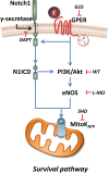
Representative scheme of intracellular pathway involved in the GPER/Notch1 cardioprotection.

Similar articles

Cited by

References

    1. Ajmani P., Yadav H. N., Singh M., Sharma P. L. (2011). Possible involvement of caveolin in attenuation of cardioprotective effect of ischemic preconditioning in diabetic rat heart. 12:43. 10.1186/1471-2261-11-43 - DOI - PMC - PubMed
    1. Boccalini G., Sassoli C., Formigli L., Bani D., Nistri S. (2015). Relaxin protects cardiac muscle cells from hypoxia/reoxygenation injury: involvement of the Notch-1 pathway. 29 239–249. 10.1096/fj.14-254854 - DOI - PubMed
    1. Bologa C. G., Revankar C. M., Young S. M., Edwards B. S., Arterburn J. B., Kiselyov A. S., et al. (2006). Virtual and biomolecular screening converge on a selective agonist for GPR30. 2 207–212. 10.1038/nchembio775 - DOI - PubMed
    1. Bopassa J. C., Eghbali M., Toro L., Stefani E. (2010). A novel estrogen receptor GPER inhibits mitochondria permeability transition pore opening and protects the heart against ischemia-reperfusion injury. 298 H16–H23. 10.1152/ajpheart.00588.2009 - DOI - PMC - PubMed
    1. Briot A., Civelek M., Seki A., Hoi K., Mack J. J., Lee S. D., et al. (2015). Endothelial NOTCH1 is suppressed by circulating lipids and antagonizes inflammation during atherosclerosis. 212 2147–2163. 10.1084/jem.20150603 - DOI - PMC - PubMed

LinkOut - more resources